
Expression of uc.189 exosomes in cervical carcinoma and its clinicopathological significance
-
摘要:目的 检测uc.189在宫颈鳞癌中的RNA表达水平,观察uc.189外泌体的数量及其与临床的病理意义。方法 应用实时荧光定量PCR(qRT-PCR)检测uc.189在58例宫颈鳞癌和癌旁正常组织中的RNA表达水平,电镜下观察宫颈鳞癌组织中uc.189外泌体的数量,并分析其与宫颈鳞癌患者病理参数的相关性。结果 uc.189在宫颈鳞癌组织中的表达水平高于癌旁正常组织,差异有统计学意义(P < 0.05)。肿瘤组织中高含量的uc.189外泌体与宫颈鳞癌的分化程度、淋巴结转移和TNM分期均密切相关(P < 0.05)。结论 uc.189可能是一种宫颈癌的新促癌基因,其可通过外泌体形式参与宫颈鳞癌的发生、发展和转移过程。Abstract:Objective To detect the RNA expression level of uc.189 in cervical squamous cell carcinoma, and to observe the number of exosomes of uc.189 as well as its clinicopathological significance.Methods Quantitative real-time polymerase chain reaction (qRT-PCR) was used to detect the expression of uc.189 RNA in 58 cases of cervical squamous cell carcinoma and adjacent normal tissues. The number of uc.189 exosomes in cervical squamous cell carcinoma was observed under electron microscope, and the correlation between the number of uc.189 exosomes and pathological parameters of cervical squamous cell carcinoma was analyzed.Results The expression level of uc.189 in the cervical squamous cell carcinoma was significantly higher than that in the adjacent normal tissues (P < 0.05). The high number of uc.189 exosomes in tumor tissue was all closely related to the differentiation degree, lymph node metastasis and TNM stage of cervical squamous cell carcinoma (P < 0.05).Conclusion The uc.189 may be a new oncogene of cervical cancer. The uc.189 promotes the occurrence, development and metastasis of cervical squamous cell carcinoma through exosome.
-
-
![]()
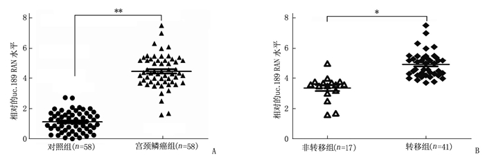
图 1 uc.189 RNA在宫颈鳞癌组织中的表达情况
A: uc.189 RNA在宫颈组织中的表达。与对照组相比, **P < 0.01。
B: uc.189 RNA在宫颈鳞癌转移组中的表达。与非转移组相比, *P < 0.05。![]()
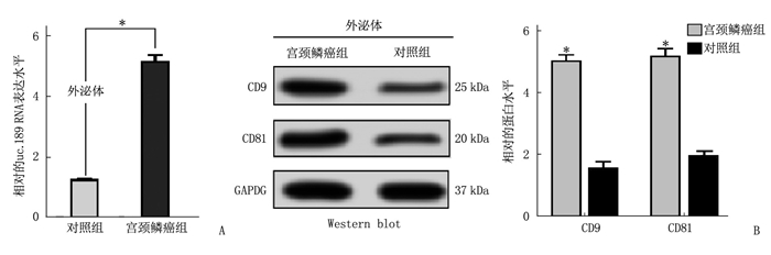
图 3 uc.189外泌体的验证
A: 对照组和宫颈癌组uc.189 RNA表达水平图; B: Western blot检测外泌体标志物图。与对照组比较, *P < 0.05。
表 1 荧光定量引物信息表
引物名称 引物序列(5′-3′) uc.189 F: GATGGTTGTACTGATGGC R: TGGTCACATGACGCTGAA U6 F: CGCTTCGGCAGCACATATAC R: TTCACGAATTTGCGTGTCAT  下载: 导出CSV
表 2 宫颈鳞癌组织中uc.189外泌体的含量及其意义(x±s)
临床病理参数 高含量组
(n=39)低含量组
(n=19)χ2 P值 患者年龄 <55岁 18 10 0.215 0.781 ≥55岁 21 9 HPV感染 有 15 5 0.834 0.397 无 24 14 肿瘤大小 <3 cm 12 11 1.988 0.252 ≥3 cm 27 8 分化程度 高~中 10 13 9.771 0.004 低 29 6 淋巴结转移 阳性 30 4 16.441 0.001 阴性 9 15 TNM分期 Ⅰ~Ⅱ 8 12 10.284 0.003 Ⅲ~Ⅳ 31 7
下载: 导出CSV
-
[1] SIEGEL R L, MILLER K D, JEMAL A. Cancer statistics, 2019[J]. CA: a Cancer J Clin, 2019, 69(1): 7-34. doi: 10.3322/caac.21551
[2] WANG L, WANG X C, LI X Y, et al. Expression of uc. 189 and its clinicopathologic significance in gynecological cancers[J]. Oncotarget, 2018, 9(7): 7453-7463. doi: 10.18632/oncotarget.23761
[3] GUO Y, WANG C H, MIAO X, et al. Upregulation of uc. 189in patients with esophageal squamous cell carcinoma and its clinicopathologic value[J]. Pathol Res Pract, 2017, 213(11): 1400-1403. doi: 10.1016/j.prp.2017.09.005
[4] ZHOU C F, MA J, HUANG L, et al. Cervical squamous cell carcinoma-secreted exosomal mi R-221-3p promotes lymphangiogenesis and lymphatic metastasis by targeting VASH1[J]. Oncogene, 2019, 38(8): 1256-1268. doi: 10.1038/s41388-018-0511-x
[5] ALENQUER M, AMORIM M J. Exosome biogenesis, regulation, and function in viral infection[J]. Viruses, 2015, 7(9): 5066-5083. doi: 10.3390/v7092862
[6] MATHIVANAN S, JI H, SIMPSON R J. Exosomes: Extracellular organelles important in intercellular communication[J]. JProteom, 2010, 73(10): 1907-1920.
[7] SIMPSON R J, LIM J W, MORITZ R L, et al. Exosomes: proteomic insights and diagnostic potential[J]. Expert Rev Proteomics, 2009, 6(3): 267-283. doi: 10.1586/epr.09.17
[8] VALADI H, EKSTRÖM K, BOSSIOS A, et al. Exosome-mediated transfer of m RNAs and micro RNAs is a novel mechanism of genetic exchange between cells[J]. Nat Cell Biol, 2007, 9(6): 654-659. doi: 10.1038/ncb1596
[9] PICCIN A, MURPHY W G, SMITH O P. Circulating microparticles: pathophysiology and clinical implications[J]. Blood Rev, 2007, 21(3): 157-171. doi: 10.1016/j.blre.2006.09.001
[10] HOSSEINI M, KHATAMIANFAR S, HASSANIAN S M, et al. Exosome-encapsulated micro RNAs as potential circulating biomarkers in colon cancer[J]. Curr Pharm Des, 2017, 23(11): 1705-1709. doi: 10.2174/1381612822666161201144634
[11] TOMASETTI M, LEE W, SANTARELLI L, et al. Exosomederived micro RNAs in cancer metabolism: possible implications in cancer diagnostics and therapy[J]. Exp Mol Med, 2017, 49(1): e285. doi: 10.1038/emm.2016.153
[12] SUNG B H, WEAVER A M. Exosome secretion promotes chemotaxis of cancer cells[J]. Cell Adh Migr, 2017, 11(2): 187-195. doi: 10.1080/19336918.2016.1273307
[13] FAN Q, YANG L, ZHANG X, et al. The emerging role of exosome-derived non-coding RNAs in cancer biology[J]. Cancer Lett, 2018, 414: 107-115. doi: 10.1016/j.canlet.2017.10.040
[14] MILANE L, SINGH A, Mattheolabakis G, et al. Exosome mediated communication within the tumor microenvironment[J]. J Control Release, 2015, 219: 278-294. doi: 10.1016/j.jconrel.2015.06.029
-
期刊类型引用(5)
1. 盘琳琳,方旭华,翟丰,桂一丁,边洲亮,陈洁. 新生儿动脉血血气分析与听力筛查结果相关性. 山东大学耳鼻喉眼学报. 2022(01): 20-24 .  百度学术
百度学术
2. 何梅,尚彪,杨雪利,鲜蓉华. 单核细胞趋化蛋白-1、可溶性细胞黏附分子-1诊断新生儿缺氧缺血性脑病的价值. 实用临床医药杂志. 2022(06): 47-50 . 本站查看
3. 黄健科. 动脉血乳酸和乳酸清除率早期评价窒息新生儿脑损伤的价值. 中国卫生标准管理. 2022(09): 60-62 . 百度学术
4. 龚晓燕,刘成博,陈燕,王利维. 尿乳酸/肌酐比值、Mb和cTnI在HIE中的表达及临床意义. 分子诊断与治疗杂志. 2022(10): 1753-1756 . 百度学术
5. 陈红兵,周瑞. 促红细胞生成素联合亚低温对缺氧缺血性脑病患儿血清因子、脑组织损伤和脑氧代谢指标的影响. 智慧健康. 2022(35): 181-185 . 百度学术
其他类型引用(0)

计量
- 文章访问数: 345
- HTML全文浏览量: 170
- PDF下载量: 8
- 被引次数: 5
 苏公网安备 32100302010246号
