Expression levels of microRNA-485-3p and Xklp2 target protein in breast cancer tissues and their clinical significance
-
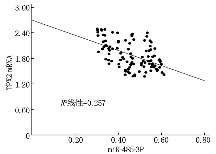
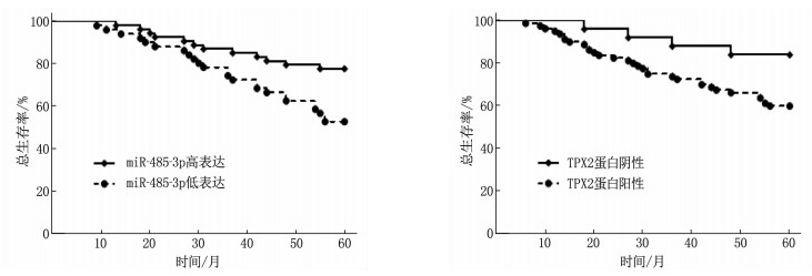
摘要:目的 探讨乳腺癌组织微小核糖核酸-485-3p(miR-485-3p)、Xklp2靶蛋白(TPX2)表达水平与患者预后的关系。方法 采用免疫组织化学法检测TPX2蛋白表达,采用实时荧光定聚合酶链反应检测miR-485-3p、TPX2 mRNA表达。采用Pearson检验分析乳腺癌组织中miR-485-3p表达与TPX2 mRNA表达的关系;随访5年,采用Kaplan-Meier法分析乳腺癌组织中miR-485-3p、TPX2蛋白表达与患者总生存率的关系,并用COX回归分析影响乳腺癌患者总生存的危险因素。结果 乳腺癌组织中TPX2蛋白阳性率、TPX2 mRNA表达水平均高于癌旁正常组织,miR-485-3p表达水平低于癌旁正常组织,差异有统计学意义(P<0.05)。乳腺癌组织中miR-485-3p、TPX2蛋白表达与患者TNM分期、淋巴结转移、Ki-67表达有关(P<0.05)。乳腺癌组织中miR-485-3p表达与TPX2 mRNA表达呈负相关(P<0.05)。miR-485-3p高表达者5年总生存率为77.78%,高于低表达者的52.94%,差异有统计学意义(P<0.05);TPX2蛋白阴性者5年总生存率为84.00%,高于阳性者的60.00%,差异有统计学意义(P<0.05)。有淋巴结转移、miR-485-3p低表达、TPX2蛋白阳性是影响乳腺癌患者总生存的独立危险因素(P<0.05)。结论 在乳腺癌组织中,miR-485-3p低表达、TPX2高表达及二者呈负相关,均可作为判断乳腺癌患者的预后指标。
-
关键词:
- 乳腺癌 /
- 微小核糖核酸-485-3p /
- Xklp2靶蛋白 /
- 预后
Abstract:Objective To investigate the relationship between the expression levels of microRNA-485-3p (miR-485-3p) and Xklp2 target protein (TPX2) in breast cancer tissues and prognosis of patients.Methods Immunohistochemistry was used to detect TPX2 protein expression, real-time fluorescent quantitative polymerase chain reaction was used to detect the expression of miR-485-3p and TPX2 mRNA. Pearson test was used to analyze the relationship between miR-485-3p expression and TPX2 mRNA expression in breast cancer tissues. Followed up for 5 years, the Kaplan-Meier method was used to analyze the relationships between the expressions of miR-485-3p, TPX2 proteins in breast cancer tissue and the overall survival rate of the patient, and COX regression analysis was used to analyze the risk factors affecting the overall survival of breast cancer patients.Results The positive rate of TPX2 protein and the level of TPX2 mRNA expression in breast cancer tissues were significantly higher than those in normal tissues adjacent to cancer, the expression level of miR-485-3p was significantly lower than that of normal tissues adjacent to cancer (P < 0.05). The expression of miR-485-3p and TPX2 proteins in breast cancer tissues were related to TNM stage, lymph node metastasis and Ki-67 expression (P < 0.05). The expression of miR-485-3p in breast cancer tissue was negatively correlated with the expression of TPX2 mRNA (P < 0.05). The 5-year overall survival rate of patients with high miR-485-3p expression was 77.78%, which was significantly higher than 52.94% of patients with low miR-485-3p expression (P < 0.05); the 5-year overall survival rate of TPX2 negative patients was 84.00%, which was significantly higher than 60.00% of positive patients (P < 0.05). The lymph node metastasis, low expression of miR-485-3p, and positive TPX2 protein were independent risk factors affecting the overall survival of breast cancer patients (P < 0.05).Conclusion In breast cancer tissues, miR-485-3p is lowly expressed and TPX2 is highly expressed. They are negatively correlated, and both can be used as prognostic indicators for judging breast cancer patients.-
Keywords:
- breast cancer /
- microRNA-485-3p /
- Xklp2 target protein /
- prognosis
-
退行性腰椎侧隐窝狭窄症(LSLRS)是由腰椎骨组织及其周围软组织退变导致无菌性炎症、神经根刺激或压迫而引发的一种疾病,临床表现为疼痛、腰部活动受限等症状。保守治疗无效的LSLRS患者应及时接受手术治疗,以解除神经根压迫症状,消除炎症[1]。椎间孔入路椎管减压术(PETD)是治疗LSLRS的常用术式,能够有效缓解神经根腹侧压迫情况,但术中显露椎弓根水平侧隐窝的操作难度较大,其对术者的技术要求较高,且该术式仅能部分开放神经根管,往往难以实现充分减压的效果[2]。经皮脊柱内镜技术因具有创伤小的优势,在腰椎滑脱症、椎间隙感染等多种腰椎疾病的治疗中发挥越来越重要的作用[3]。全内镜下椎板开窗术(Endo-LOVE)已被应用于LSLRS的治疗中,有助于实现腰椎管的充分减压[4]。本研究比较Endo-LOVE与PETD对LSLRS患者的近期疗效及并发症发生情况,现将结果报告如下。
1. 资料与方法
1.1 一般资料
选取2019年3月—2022年11月北京中医药大学东直门医院收治的160例LSLRS患者作为研究对象,采用随机数字表法分为对照组和观察组,每组80例。纳入标准; ①符合《退变性腰椎管狭窄症临床诊疗指南》[5]中的LSLRS诊断标准(主要表现为神经源性间歇性跛行; 长期存在坐骨神经痛; 神经支配区域出现皮肤感觉减退、肌力下降及肌腱反射减弱; 影像学检查显示中央管无明显狭窄,仅有腰椎侧隐窝狭窄)者; ②近3个月内未参加其他临床试验者; ③年龄≥60岁者; ④可耐受手术,且术后可完成随访者; ⑤签署知情同意书者。排除标准; ①多节段病变者; ②单纯腰椎间盘突出症,或有马尾综合征者; ③伴有腰部骨折、脊柱感染、心脑血管疾病者; ④伴有先天性脊柱畸形者; ⑤伴有血液系统疾病者; ⑥伴有严重骨质疏松者。2组患者一般资料比较,差异无统计学意义(P>0.05), 具有可比性,见表 1。
表 1 2组患者一般资料比较(x±s)[n(%)]一般资料 分类 对照组(n=80) 观察组(n=80) χ2/t P 性别 男 48(60.00) 44(55.00) 0.409 0.522 女 32(40.00) 36(45.00) 年龄/岁 70.95±7.53 69.87±7.86 0.887 0.376 责任节段 L2~3节段 4(5.00) 2(2.50) 1.690 0.639 L3~4节段 15(18.75) 18(22.50) L4~5节段 55(68.75) 51(63.75) L5~S1节段 6(7.50) 9(11.25) 病程 < 6个月 24(30.00) 21(26.25) 0.309 0.857 6~12个月 36(45.00) 37(46.25) >12个月 20(25.00) 22(27.50) 内科合并疾病 高血压 34(42.50) 38(47.50) 1.237 0.266 冠心病 21(26.25) 18(22.50) 2.884 0.089 脑卒中 12(15.00) 16(20.00) 0.291 0.589 糖尿病 17(21.25) 20(25.00) 0.572 0.449 1.2 方法
对照组接受PETD治疗。患者取俯卧位,头部、胸部及双髂部垫软枕,确保腹部悬空。利用C臂机进行定位,在病椎的椎间隙水平线和后正中线上做标记。局部麻醉后,通过1 cm切口,在导丝引导下置入工作套管。透视下确认套管位置无误后,使用椎间孔镜进行手术,切除部分骨质,扩大椎间孔,必要时修整黄韧带和椎间盘组织。手术成功的标志是扩大的骨性空间中无组织突出,神经根活动自如。最后进行止血处理,缝合切口,完成手术。
观察组接受Endo-LOVE治疗。患者的体位和麻醉方法与对照组相同。利用C臂机进行定位,于病椎间隙正中线旁开1.0~1.5 cm处进针。经皮肤和筋膜切开后,插入工具以扩张软组织,并置入工作套管至椎板间隙。使用椎间孔镜观察并清理术区,切开黄韧带和部分骨质,以显露并扩大椎板间隙。通过调整套管位置并切除骨质和黄韧带,解除对神经结构的压迫,并移除突出组织。手术成功的标志是神经根和硬膜囊能够自主搏动。最后进行止血处理,缝合切口,完成手术。
2组患者术后均接受严格的心电监护,至少卧床休息24 h, 并在指导下进行踝泵训练。24 h后,若患者无明显疼痛感,可在佩戴腰围的情况下离床活动。
1.3 观察指标
① 手术相关指标,记录2组患者的术中出血量、手术时间和住院时间。②并发症发生情况,观察2组患者下肢麻木、发热、术后腰背痛和血管神经损伤的发生情况。③疗效,随访6个月,评估2组患者的疗效,包括优(无痛,活动不受限,原有症状无残留)、良(轻微疼痛、麻木,能够参加适当调整后的工作,下肢功能轻度受限)、可(疼痛、麻木改善,但对参加工作存在一定影响,下肢功能受限)、差(持续性疼痛、麻木,严重影响参加工作)。④骨性侧隐窝角和软性侧隐窝角, 2组患者于术前及术后72 h分别接受腰椎磁共振成像(MRI)或CT检查,在水平位影像上测量冠状面和半矢状面上构成侧隐窝两边的夹角,分别定义为骨性侧隐窝角和软性侧隐窝角。⑤ Oswestry功能障碍指数(ODI)评分[6], 该评分系统共包含10个方面,总分为100分,评分越高表示病情和功能障碍越严重。⑥疼痛评分,采用视觉模拟评分法(VAS)[7]进行评估,评分范围为0~10分, 0分表示无痛, 10分表示疼痛无法忍受,评分与疼痛程度呈正相关。⑦日本骨科协会腰痛疾病评定表(JOA)评分[8], 该量表包含4个维度,总分为29分,评分越高表示功能性障碍越轻。
1.4 统计学分析
采用SPSS 26.0统计学软件分析数据,符合正态分布并满足方差齐性的计量资料以(x±s)表示,组间比较采用独立样本t检验,组内比较采用配对样本t检验,计数资料以[n(%)]描述,组间比较采用χ2检验。所有统计检验以α=0.05为显著性水平标准,P < 0.05为差异有统计学意义。
2. 结果
2.1 手术相关指标和并发症发生情况比较
观察组术中出血量少于对照组,手术时间、住院时间短于对照组,差异有统计学意义(P < 0.05); 观察组下肢麻木、发热、术后腰背痛等并发症发生率与对照组比较,差异无统计学意义(P>0.05), 见表 2。
表 2 2组患者手术相关指标和并发症发生情况比较(x±s)[n(%)]指标 分类 对照组(n=80) 观察组(n=80) 手术相关指标 术中出血量/mL 13.66±3.12 9.15±2.55* 手术时间/min 73.63±10.74 68.02±9.11* 住院时间/d 7.58±1.36 6.41±1.08* 并发症 下肢麻木 4(5.00) 3(3.75) 发热 2(2.50) 1(1.25) 术后腰背痛 1(1.25) 1(1.25) 血管神经损伤 0 0 合计 7(8.75) 4(5.00) 与对照组比较, *P < 0.05。 2.2 骨性侧隐窝角和软性侧隐窝角比较
术前, 2组骨性侧隐窝角、软性侧隐窝角比较,差异无统计学意义(P>0.05); 术后72 h, 2组骨性侧隐窝角、软性侧隐窝角均较术前增大,且观察组大于对照组,差异有统计学意义(P < 0.05), 见表 3。
表 3 2组患者手术前后骨性侧隐窝角和软性侧隐窝角比较(x±s)° 组别 软性侧隐窝角 骨性侧隐窝角 术前 术后72 h 术前 术后72 h 对照组(n=80) 13.52±3.58 24.87±4.95* 17.41±5.02 29.86±5.22* 观察组(n=80) 13.43±3.96 30.26±5.57*# 17.23±4.86 35.14±6.07*# 与术前比较, *P < 0.05; 与对照组比较, #P < 0.05。 2.3 疼痛评分、ODI评分和JOA评分比较
术前, 2组疼痛评分、ODI评分和JOA评分比较,差异均无统计学意义(P>0.05); 术后3个月时, 2组JOA评分均较术前升高,疼痛评分、ODI评分均较术前降低,且观察组JOA评分高于对照组,疼痛评分、ODI评分低于对照组,差异有统计学意义(P < 0.05), 见表 4。
表 4 2组患者手术前后疼痛评分、ODI评分和JOA评分比较(x±s)分 组别 疼痛评分 ODI评分 JOA评分 术前 术后3个月时 术前 术后3个月时 术前 术后3个月时 对照组(n=80) 6.52±1.47 1.87±0.56* 75.52±10.05 31.23±8.05* 15.56±4.04 23.74±4.16* 观察组(n=80) 6.47±1.53 1.21±0.43*# 76.41±11.53 21.77±6.53*# 15.37±3.96 27.08±4.39*# ODI; Oswestry功能障碍指数; JOA; 日本骨科协会腰痛疾病评定表。与术前比较, *P < 0.05; 与对照组比较, #P < 0.05。 2.4 疗效比较
观察组优良率为96.25%(77/80), 高于对照组的87.50%(70/80), 差异有统计学意义(P < 0.05), 见表 5。
表 5 2组患者疗效比较[n(%)]组别 优 良 可 差 总优良 对照组(n=80) 36(45.00) 34(42.50) 10(12.50) 0 70(87.50) 观察组(n=80) 58(72.50) 19(23.75) 3(3.75) 0 77(96.25)* 与对照组比较, *P < 0.05。 2.5 典型病例分析
典型病例1: 患者男, 55岁,主诉腰痛伴右侧小腿麻木1年,保守治疗3个月无效,入院后接受PETD治疗。术前影像学检查显示L4~5椎间盘突出,椎间孔及侧隐窝处狭窄,术前CT轴位图像显示L5侧隐窝处存在骨性狭窄,对L5神经根产生压迫,故术中需进行侧隐窝减压处理。PETD治疗时,选择L4~5右侧入路进行手术。术后,患者右下肢疼痛VAS评分从术前的7分降至1分,症状显著缓解,手术效果满意。该患者手术前后影像学检查结果见图 1。
典型病例2: 患者女, 42岁,主诉腰痛伴左下肢疼痛2年,保守治疗3个月无效,入院后接受Endo-LOVE治疗。术前MRI及CT检查结果显示, L4~5椎间盘突出继发椎间孔及侧隐窝区域狭窄,术前轴位图像显示右侧侧隐窝区域存在骨性狭窄,术中需探查明确后进行有效的侧隐窝区域减压。术后复查轴位CT, 可见侧隐窝角较术前增大。术后,患者症状显著缓解,下肢疼痛VAS评分由术前的8分降至0分,手术效果满意。该患者手术前后影像学检查结果见图 2。
3. 讨论
LSLRS好发于老年人,多由退行性变引起,早期起病隐匿,随着病程进展可能累及多个椎体,导致穿过硬膜囊的神经根在侧隐窝狭窄处受到压迫。LSLRS患者常表现出疼痛、麻木、间歇性跛行等症状,严重影响正常生活和工作[9]。随着人口老龄化的加剧, LSLRS的发生率呈现逐渐升高趋势[10]。目前, LSLRS的治疗方案主要包括保守治疗和手术治疗。保守治疗涵盖功能训练、物理治疗及镇痛药物治疗等,若保守治疗无效,患者需接受手术治疗,以解除神经根管压迫,缓解疼痛、麻木、间歇性跛行等症状[11]。在治疗LSLRS的多种术式中, Endo-LOVE与PETD在临床实践中较为常用。朱凯等[12]研究认为,采用Endo-LOVE方案治疗LSLRS能够充分扩大侧隐窝,取得良好的临床疗效。
骨性侧隐窝角和软性侧隐窝角是评估侧隐窝狭窄程度的关键指标, JOA评分、疼痛评分及ODI评分均为评价LSLRS病情严重程度的常用工具。本研究结果显示,术后72 h, 2组患者的骨性侧隐窝角和软性侧隐窝角均较术前显著增大,且观察组显著大于对照组。由此表明, Endo-LOVE方案对LSLRS具有良好的近期疗效,能够有效改善侧隐窝狭窄状况,并减轻腰椎功能障碍和疼痛症状。尽管PETD可通过椎间孔的安全三角区进行手术,移除突出组织,并部分切除黄韧带和骨组织以缓解神经根管压迫,但其对神经根的减压效果有限。值得注意的是,高髂嵴或横突阻挡会增加穿刺难度,手术操作时应予以重视[13]。Endo-LOVE能够更充分地扩大侧隐窝,彻底开放神经根管,增大骨性侧隐窝角及软性侧隐窝角,一次性解决导致侧隐窝狭窄的因素,因此疗效更佳。使用环锯切除椎板、小关节骨质及黄韧带,可有效缓解神经根压力,摘除压迫性组织,减少神经损伤,显著减轻患者疼痛,促进术后康复[14]。
本研究中,与对照组比较,观察组术中出血量更少,手术时间和住院时间更短,但2组下肢麻木、发热、术后腰背痛等并发症发生率无显著差异。由此提示, Endo-LOVE方案应用于LSLRS患者的治疗中,具有手术时间短、术中出血量少、术后恢复快等优势。Endo-LOVE技术的穿刺路径较短,术中咬除侧隐窝背侧骨质的操作更为便捷,从而缩短了手术时间。此外, Endo-LOVE术中对软组织的剥离较少,减少了出血和创伤,从而减轻了患者术后的疼痛感,使其能更快开始康复训练,加速康复进程,缩短住院时间[15]。理论上,出血量减少、手术时间缩短可以减轻手术创伤,缩短术中辐射暴露时间,进而降低术后并发症发生风险。然而,本研究中2组患者的并发症发生率无显著差异,这可能与样本量较少引起的统计偏倚有关,未来需开展大样本进一步探讨。值得注意的是, Endo-LOVE与PETD均需在C臂机下定位操作,可能引起放射性损伤,因此在操作过程中应注意提高精确度,尽量缩短患者的射线暴露时间。
综上所述,相较于PETD方案, Endo-LOVE方案治疗LSLRS具有手术时间短、术中出血量少、近期疗效好的优点。
-
表 1 乳腺癌组织和癌旁正常组织中TPX2蛋白表达情况
组别 n TPX2蛋白 阳性 阴性 阳性率/% 癌旁正常组织 105 39 66 37.14 乳腺癌组织 105 80 25 76.19* 与癌旁正常组织比较, * P<0.05。 表 2 乳腺癌组织和癌旁正常组织中miR-485-3p、TPX2 mRNA表达水平比较(x±s)
组别 n miR-485-3p TPX2 mRNA 癌旁正常组织 105 1.00±0.27 1.01±0.26 乳腺癌组织 105 0.45±0.12* 1.83±0.49* 与癌旁正常组织比较, * P<0.05。 表 3 乳腺癌组织中miR-485-3p、TPX2蛋白表达与临床病理特征的关系[n(%)]
临床病理特征 n miR-485-3p χ2值 P值 TPX2蛋白 χ2值 P值 低表达(n=51) 高表达(n=54) 阳性(n=80) 阴性(n=25) 年龄 <48岁 49 22(44.90) 27(55.10) 0.496 0.481 35(71.43) 14(28.57) 1.148 0.284 ≥48岁 56 29(51.79) 27(48.21) 45(80.36) 11(19.64) 肿瘤直径 <2 cm 47 19(40.43) 28(59.57) 2.260 0.133 32(68.09) 15(31.91) 3.081 0.079 ≥2 cm 58 32(55.17) 26(44.83) 48(82.76) 10(17.24) TNM分期 Ⅰ+Ⅱ期 71 28(39.44) 43(60.56) 7.325 0.007 49(69.01) 22(30.99) 6.225 0.013 Ⅲ期 34 23(67.65) 11(32.35) 31(91.18) 3(8.82) 淋巴结转移 有 45 30(66.67) 15(33.33) 10.323 0.001 39(86.67) 6(13.33) 4.764 0.029 无 60 21(35.00) 39(65.00) 41(68.33) 19(31.67) PR表达 阴性 53 27(50.94) 26(49.06) 0.241 0.623 43(81.13) 10(18.87) 1.441 0.230 阳性 52 24(46.15) 28(53.85) 37(71.15) 15(28.85) ER表达 阴性 76 39(51.32) 37(48.68) 0.830 0.362 59(77.63) 17(22.37) 0.315 0.575 阳性 29 12(41.38) 17(58.62) 21(72.41) 8(27.59) HER-2表达 阴性 57 28(49.12) 29(50.88) 0.015 0.902 44(77.19) 13(22.81) 0.069 0.793 阳性 48 23(47.92) 25(52.08) 36(75.00) 12(25.00) Ki-67表达 阴性 44 14(31.82) 30(68.18) 8.510 0.004 28(63.64) 16(36.36) 6.580 0.010 阳性 61 37(60.66) 24(39.34) 52(85.25) 9(14.75) 表 4 影响乳腺癌患者总生存的危险因素
变量 单因素分析 多因素分析 HR值 95% CI P值 HR值 95% CI P值 年龄(≥48岁/<48岁) 1.060 0.821~1.369 0.075 — — — 肿瘤直径(≥2 cm/<2 cm) 0.944 0.774~1.152 1.002 — — — TNM分期(Ⅲ期/Ⅰ+Ⅱ期) 1.589 1.260~2.004 0.004 1.144 0.867~1.509 0.069 淋巴结转移(有/无) 1.680 1.418~1.990 0.000 1.421 1.245~1.623 0.030 PR表达(阴性/阳性) 0.873 0.643~1.185 1.197 — — — ER表达(阴性/阳性) 0.993 0.725~1.361 0.082 — — — HER-2表达(阴性/阳性) 0.995 0.698~1.417 0.081 — — — Ki-67表达(阳性/阴性) 1.580 1.294~1.930 0.006 1.197 0.912~1.570 0.058 miR-485-3p(低表达/高表达) 1.835 1.371~2.455 0.000 1.568 1.163~2.114 0.013 TPX2蛋白(阳性/阴性) 1.517 1.083~2.126 0.018 1.399 1.046~1.872 0.037 -
[1] VERRET B, SOURISSEAU T, STEFANOVSKA B, et al. The influence of cancer molecular subtypes and treatment on the mutation spectrum in metastatic breast cancers[J]. Cancer Res, 2020, 80(15): 3062-3069. doi: 10.1158/0008-5472.CAN-19-3260
[2] 马跃, 高英静, 何浪. 乳腺癌差异表达miRNA在预后中的意义[J]. 医学研究杂志, 2018, 47(1): 39-44. https://www.cnki.com.cn/Article/CJFDTOTAL-YXYZ201801013.htm [3] 艾尼·沙塔尔, 闫焕英, 丁伟, 等. 长链非编码RNA MALAT1靶向miR-485-3p调控乳腺癌细胞对紫杉醇的耐药性[J]. 南方医科大学学报, 2020, 40(5): 698-702. https://www.cnki.com.cn/Article/CJFDTOTAL-DYJD202005014.htm [4] JIANG Y Q, LIU Y, TAN X L, et al. TPX2 as a novel prognostic indicator and promising therapeutic target in triple-negative breast cancer[J]. Clin Breast Cancer, 2019, 19(6): 450-455. doi: 10.1016/j.clbc.2019.05.012
[5] TAHERDANGKOO K, KAZEMINEZHAD S R, HAJJARI M R, et al. miR-485-3p suppresses colorectal cancer via targeting TPX2[J]. Bratislava Med J, 2020, 121(4): 302-307. doi: 10.4149/BLL_2020_048
[6] 中国抗癌协会乳腺癌专业委员会. 中国抗癌协会乳腺癌诊治指南与规范(2017年版)[J]. 中国癌症杂志, 2017, 27(9): 695-759. https://www.cnki.com.cn/Article/CJFDTOTAL-ZGAZ201709004.htm [7] WANG Y W, ZHAO S, YUAN X Y, et al. miR-4732-5p promotes breast cancer progression by targeting TSPAN13[J]. J Cell Mol Med, 2019, 23(4): 2549-2557. doi: 10.1111/jcmm.14145
[8] 许发功, 杨立. 乳腺癌组织中miR-144-3p和SGK3的表达水平及临床意义[J]. 中国临床研究, 2019, 32(2): 173-178. https://www.cnki.com.cn/Article/CJFDTOTAL-ZGCK201902008.htm [9] LAI J J, XIN J, FU C D, et al. CircHIPK3 promotes proliferation and metastasis and inhibits apoptosis of renal cancer cells by inhibiting MiR-485-3p[J]. Cancer Cell Int, 2020, 20: 248. doi: 10.1186/s12935-020-01319-3
[10] WANG M, CAI W R, MENG R, et al. miR-485-5p suppresses breast cancer progression and chemosensitivity by targeting survivin[J]. Biochem Biophys Res Commun, 2018, 501(1): 48-54. doi: 10.1016/j.bbrc.2018.04.129
[11] WANG X, ZHOU X, ZENG F, et al. miR-485-5p inhibits the progression of breast cancer cells by negatively regulating MUC1[J]. Breast Cancer, 2020, 27(4): 765-775. doi: 10.1007/s12282-020-01075-2
[12] 王庆亮, 赵艳峰, 蒋燕, 等. TPX2与肿瘤关系的研究进展[J]. 实用肿瘤学杂志, 2021, 35(1): 83-86. https://www.cnki.com.cn/Article/CJFDTOTAL-SYZL202101022.htm [13] JIANG T C, SUI D M, YOU D, et al. RETRACTED ARTICLE: MiR-29a-5p inhibits proliferation and invasion and induces apoptosis in endometrial carcinoma via targeting TPX2[J]. Cell Cycle, 2018, 17(10): 1268-1278. doi: 10.1080/15384101.2018.1475829
[14] ZOU J, HUANG R Y, JIANG F N, et al. Overexpression of TPX2 is associated with progression and prognosis of prostate cancer[J]. Oncol Lett, 2018, 16(3): 2823-2832. http://www.ingentaconnect.com/content/sp/ol/2018/00000016/00000003/art00008
[15] ZHOU F, WANG M, AIBAIDULA M, et al. TPX2 promotes metastasis and serves as a marker of poor prognosis in non-small cell lung cancer[J]. Med Sci Monit, 2020, 26: e925147. https://pubmed.ncbi.nlm.nih.gov/32748897/#:~:text=TPX2%20Promotes%20Metastasis%20and%20Serves%20as%20a%20Marker,mechanisms%20would%20aid%20in%20initiating%20effective%20clinical%20treatment.
[16] TAN G Z, LI M, TAN X, et al. MiR-491 suppresses migration and invasion via directly targeting TPX2 in breast cancer[J]. Eur Rev Med Pharmacol Sci, 2019, 23(22): 9996-10004. http://www.ncbi.nlm.nih.gov/pubmed/31799669


 下载:
下载:


 苏公网安备 32100302010246号
