
Effect observation of 3D printing adjuvant therapy in treatment of patients with posterior Pilon fracture after treatment
-
摘要:目的
观察3D打印辅助治疗后Pilon骨折的临床疗效。
方法回顾性分析2012年1月—2020年12月溧阳市人民医院骨科收治的92例后Pilon骨折患者的临床资料,其中术前应用3D打印模拟手术的47例患者设为3D打印组,未应用3D打印模拟手术的45例患者设为对照组。记录2组患者的手术相关并发症,比较2组患者手术时间、术中X线透视次数、手术复位情况、骨折临床愈合时间、末次随访时美国足踝外科协会(AOFAS)足踝评分系统评分及踝关节疼痛视觉模拟评分法(VAS)评分。
结果所有患者均获12~20个月随访。所有患者术后切口均达到Ⅰ期愈合,无患者发生切口感染、皮肤坏死、下肢深静脉血栓、足趾马缰绳畸形等并发症。3D打印组手术时间、术中透视次数分别为(81.4±9.4) min、(13.0±2.3)次,对照组分别为(94.9±11.6) min、(18.4±3.2)次,差异有统计学意义(P < 0.05)。对照组有3例患者后踝骨折复位不良, 3D打印组无患者发生后踝骨折复位不良,差异无统计学意义(P>0.05)。3D打印组骨折愈合时间、术后AOFAS评分、VAS评分分别为(12.9±1.0)周、(92.8±4.2)分、(1.1±0.8)分,对照组分别为(13.2±1.3)周、(90.8±4.5)分、(1.3±0.8)分,差异无统计学意义(P>0.05)。
结论应用3D打印技术辅助治疗可以优化后Pilon骨折手术方案,缩短手术时间,减少术中透视次数,实现个体化精准治疗。
Abstract:ObjectiveTo observe the clinical effect of 3D printing adjuvant therapy in treatment of patients with posterior Pilon fracture after treatment.
MethodsThe clinical materials of 92 patients with posterior Pilon fracture from January 2012 to December 2020 in the Department of Orthopedics of Liyang People′s Hospital were retrospectively analyzed. Among them, 47 patients with preoperative 3D printing for simulating operation were selected as 3D printing group, and 45 patients without 3D printing for simulating operation were selected as control group. The operation related complications were recorded in both groups, and the operation time, intraoperative X-ray fluoroscopy times, condition of surgical reduction, clinical healing time of fractures, the score of ankle scoring system of the American College of Foot and Ankle Surgeons (AOFAS) and the score of Visual Analogue Scale (VAS) of ankle pain at the last follow-up were compared between the two groups.
ResultsAll the patients were followed up for 12 to 20 months. All the patients achieved primary healing after operation, and no patient had complications such as incision infection, skin necrosis, deep venous thrombosis of lower limbs, and checkrein deformity of toes. The operation time and intraoperative fluoroscopy times in the 3D printing group were (81.4±9.4) min and (13.0±2.3) times respectively, which were significantly shorter and less than (94.9±11.6) min and (18.4±3.2) times in the control group (P < 0.05). In the control group, there were 3 cases with poor reduction of posterior malleolus fracture, but there were no patients with poor reduction of posterior malleolus fracture in the 3D printing group, and there was no significant difference between two groups (P>0.05). The fracture healing time, postoperative AOFAS score and VAS score in the 3D printing group were (12.9±1.0) weeks, (92.8±4.2) points and (1.1±0.8) points respectively, while were (13.2±1.3) weeks, (90.8±4.5) points and (1.3±0.8) points respectively in the control group, and there were no significant differences between two groups (P>0.05).
ConclusionApplication of 3D printing adjuvant therapy can optimize the operation plan of posterior Pilon fracture, shorten the operation time, reduce the number of intraoperative fluoroscopy, and achieve individualized precision treatment.
-
-
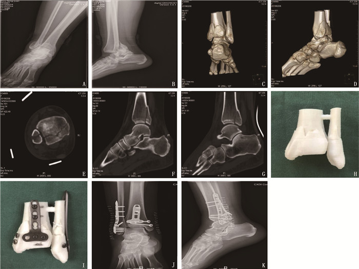
![]()
图 1 1例42岁右侧后Pilon骨折(Klammer Ⅱ型)女性患者的X线片和3D打印模型
A、B: 术前踝关节正侧位X线片显示Klammer Ⅱ型后Pilon骨折; C、D: 术前踝关节CT三维重建显示后踝骨折延续至内踝; E、F、G: 踝关节CT平扫与后内侧、后外侧矢状位重建; H、I: 3D打印模型及术前模拟情况,并确定骨折复位标记、接骨板位置、螺钉位置及长度; J、K: 术后踝关节正侧位X线片显示骨折解剖复位,内固定位置良好。
表 1 2组后Pilon骨折患者一般资料比较(x±s)
组别 n 性别 年龄/岁 受伤部位 致伤原因 Klammer分型 术前住院时间/d 男 女 左侧 右侧 交通伤 坠落伤 扭伤 Ⅰ型 Ⅱ型 Ⅲ型 对照组 45 22 23 51.3±11.7 23 22 18 13 14 18 13 14 8.8±1.7 3D打印组 47 22 25 52.0±12.8 23 24 17 14 16 20 12 15 8.9±1.7  下载: 导出CSV
表 2 2组患者手术相关指标及术后随访情况比较(x±s)[n(%)]
组别 n 随访时间/月 手术时间/min 术中透视次数/次 复位不良 骨折愈合时间/周 AOFAS评分/分 VAS评分/分 对照组 45 16.5±2.1 94.9±11.6 18.4±3.2 3(6.7) 13.2±1.3 90.8±4.5 1.3±0.8 3D打印组 47 16.6±1.6 81.4±9.4* 13.0±2.3* 0 12.9±1.0 92.8±4.2 1.1±0.8 AOFAS: 美国足踝外科协会; VAS: 视觉模拟评分法。与对照组比较, *P < 0.05。
下载: 导出CSV
-
[1] HANSEN S. Functional reconstruction of the foot and ankle[M]. Philadelphia: Lippincott Williams & Wilkins, 2000: 37-46.
[2] 秦晓东, 张宇, 方永详, 等. 过度跖屈型踝关节骨折的初步探讨[J]. 中华创伤骨科杂志, 2017, 19(12): 1029-1035. doi: 10.3760/cma.j.issn.1671-7600.2017.12.005 [3] ZHANG J Z, WANG H, PEN C, et al. Characteristics and proposed classification system of posterior pilon fractures[J]. Medicine, 2019, 98(3): e14133. doi: 10.1097/MD.0000000000014133
[4] WANG J W, WANG X Y, XIE L Z, et al. Comparison of radiographs and CT features between posterior Pilon fracture and posterior malleolus fracture: a retrospective cohort study[J]. Br J Radiol, 2020, 93(1110): 20191030. doi: 10.1259/bjr.20191030
[5] CHAPARRO F, AHUMADA X, URBINA C, et al. Posterior pilon fracture: Epidemiology and surgical technique[J]. Injury, 2019, 50(12): 2312-2317. doi: 10.1016/j.injury.2019.10.007
[6] VACAS-SÁNCHEZ E, OLAYA-GONZÁLEZ C, ABARQUERO-DIEZHANDINO A, et al. How to address the posterior malleolus in ankle fractures A decision-making model based on the computerised tomography findings[J]. Int Orthop, 2020, 44(6): 1177-1185. doi: 10.1007/s00264-020-04481-5
[7] 刘春光, 宋朋飞, 李兴华. 3D打印在治疗后Pilon骨折中的应用[J]. 中华实验外科杂志, 2020, 37(4): 632-634. doi: 10.3760/cma.j.cn421213-20190720-01017 [8] KLAMMER G, KADAKIA A R, JOOS D A, et al. Posterior pilon fractures: a retrospective case series and proposed classification system[J]. Foot Ankle Int, 2013, 34(2): 189-199. doi: 10.1177/1071100712469334
[9] 王旭, 耿翔, 张超, 等. 后pilon骨折Die-punch骨块的CT分型及应用[J]. 中华创伤骨科杂志, 2018, 20(6): 470-475. doi: 10.3760/cma.j.issn.1671-7600.2018.06.003 [10] 刘波, 乔之军, 曹光华, 等. 后内侧胫后肌腱前方入路联合后外侧入路切开复位内固定治疗Klammer Ⅱ/Ⅲ型后pilon样骨折[J]. 中华创伤杂志, 2021, 37(12): 1099-1104. doi: 10.3760/cma.j.cn501098-20210408-00232 [11] 张宇, 孙海钰, 陈斌. 后外侧入路在后Pilon骨折中的应用[J]. 实用骨科杂志, 2019, 25(5): 438-441. https://www.cnki.com.cn/Article/CJFDTOTAL-SGKZ201905014.htm [12] CAMPBELL S T, DEBAUN M R, KLEWENO C P, et al. Simultaneous posterolateral and posteromedial approaches for fractures of the entire posterior tibial plafond: a safe technique for effective reduction and fixation[J]. J Orthop Trauma, 2022, 36(1): 49-53. doi: 10.1097/BOT.0000000000002144
[13] ZBEDA R M, FRIEDEL S P, KATCHIS S D, et al. Open reduction and internal fixation of posterior malleolus fractures via a posteromedial approach[J]. Orthopedics, 2020, 43(3): e166-e170.
[14] SUKUR E, AKMAN Y E, GOKCEN H B, et al. Open reduction in pilon variant posterior malleolar fractures: Radiological and clinical evaluation[J]. Orthop Traumatol Surg Res, 2017, 103(5): 703-707. doi: 10.1016/j.otsr.2017.05.012
[15] MARTIN K D, TRIPP C T, HUH J. Outcomes of posterior arthroscopic reduction and internal fixation (PARIF) for the posterior malleolar fragment in trimalleolar ankle fractures[J]. Foot Ankle Int, 2021, 42(2): 157-165. doi: 10.1177/1071100720955149
[16] LIU T, CHENG Y H, QU W Q. A fibular Notch approach for the treatment of ankle fractures involving the distal tibial plafond[J]. J Orthop Surg Res, 2021, 16(1): 120. doi: 10.1186/s13018-021-02270-3
[17] KIM Y J, LEE J H. Posterior inferior tibiofibular ligament release to achieve anatomic reduction of posterior malleolar fractures[J]. J Foot Ankle Surg, 2018, 57(1): 86-90. doi: 10.1053/j.jfas.2017.08.012
[18] SHIWAKU K, TERAMOTO A, IBA K, et al. The prevalence of posterior inferior tibiofibular ligament and inferior tibiofibular transverse ligament injuries in syndesmosis-injured ankles evaluated by oblique axial magnetic resonance imaging: a retrospective study[J]. BMC Musculoskelet Disord, 2022, 23(1): 264. doi: 10.1186/s12891-022-05220-0
[19] MARTIN K D. Posterior arthroscopic reduction and internal fixation for treatment of posterior malleolus fractures[J]. Foot Ankle Int, 2020, 41(1): 115-120. doi: 10.1177/1071100719891978
[20] JIANG Z, ZHANG C, QIN J J, et al. Posterior pilon fracture treated by opening the Fibula fracture gap[J]. J Orthop Surg Res, 2022, 17(1): 214. doi: 10.1186/s13018-022-03106-4
[21] 陈东亮, 郑良孝, 朱朝辉, 等. 骨折间隙直视下复位固定后踝移位骨折[J]. 中国矫形外科杂志, 2020, 28(2): 177-181. https://www.cnki.com.cn/Article/CJFDTOTAL-ZJXS202002022.htm [22] SUN C G, PENG X Q, FEI Z G, et al. The CT morphological characteristics and the clinical management strategy of posterior malleolar fractures with talar subluxation[J]. Am J Transl Res, 2021, 13(6): 6478-6487.
[23] 衡科, 陶涛, 魏辉, 等. 不同内固定方式联合入路治疗Klammer Ⅲ型后pilon骨折效果[J]. 实用临床医药杂志, 2021, 25(12): 66-69. doi: 10.7619/jcmp.20210822 [24] GAO M F, LIU N C, CHENG Y, et al. Treatment outcomes of the posterolateral approach of plate fixation for posterior pilon fractures[J]. Exp Ther Med, 2019, 17(5): 4267-4272.
-
期刊类型引用(4)
1. 殷华芳,沙莎,蔡依玲,于波,刘佳,何佳,孙玲娣,王坚. 宫颈癌放射治疗相关卵巢损伤的分子机制及防治策略研究进展. 实用临床医药杂志. 2024(10): 141-144 .  本站查看
本站查看
2. 张玉洲,李晓敏,孙少霖. 蓝萼乙素通过Akt/BAD通路对宫颈癌裸鼠移植瘤生长的影响. 现代药物与临床. 2024(06): 1384-1389 . 百度学术
3. 沈静,张丽华,徐晶晶,吕萌萌,吴东辰. 卡瑞利珠单抗联合白蛋白结合型紫杉醇对晚期宫颈癌患者肿瘤标志物、免疫功能和血管新生指标的影响. 现代生物医学进展. 2024(13): 2592-2595 . 百度学术
4. 曾美男. 早期宫颈癌术后不同治疗方法的临床效果分析. 实用妇科内分泌电子杂志. 2023(24): 28-30+85 . 百度学术
其他类型引用(0)
计量
- 文章访问数: 166
- HTML全文浏览量: 64
- PDF下载量: 12
- 被引次数: 4
 苏公网安备 32100302010246号