Experimental study of neurotrophin-3 overexpression induced pluripotent stem cells in treating rats with diabetes-induced erectile dysfunction
-
摘要:目的
探讨神经营养素-3过表达诱导多能干细胞(iPSC-NT-3)对糖尿病性勃起功能障碍(DIED)大鼠的治疗作用。
方法腹腔注射链脲佐菌素建立糖尿病模型大鼠,皮下注射阿扑吗啡对DIED模型进行验证。选择24只造模成功的DIED大鼠,随机分为模型对照组、诱导多能干细胞(iPSC)治疗组和iPSC-NT-3治疗组; 腹腔注射柠檬酸钠-柠檬酸缓冲液的8只SD大鼠作为正常对照组。各组大鼠腹腔注射戊巴比妥钠进行麻醉, iPSC治疗组和iPSC-NT-3治疗组分别将iPSC和iPSC-NT-3用微量注射针注射入大鼠阴茎海绵体内,正常对照组和模型对照组同法注射同体积的磷酸缓冲液。治疗后第4周,观察各组大鼠的一般情况,评估性功能和阴茎勃起功能。采用定量聚合酶链反应(q-PCR)和Western blot实验检测神经营养素-3(NT-3)、CD31、血管内皮生长因子(VEGF)、内皮型一氧化氮合酶(eNOS)、结蛋白(Desmin)、α-平滑肌肌动蛋白(α-SMA)的mRNA和蛋白的表达。
结果治疗前和治疗后4周,模型对照组、iPSC治疗组和iPSC-NT-3治疗组大鼠体质量和血糖水平的组内、组间差异均无统计学意义(P>0.05)。在大鼠首次爬背时间比较中,模型对照组较正常对照组延长, iPSC治疗组、iPSC-NT-3治疗组较模型对照组缩短, iPSC-NT-3治疗组较iPSC治疗组缩短,差异均有统计学意义(P < 0.05); 在舔嗅次数、骑跨次数和插入次数的比较中,模型对照组均较正常对照组减少, iPSC治疗组、iPSC-NT-3治疗组较模型对照组增加, iPSC-NT-3治疗组较iPSC治疗组增加,差异均有统计学意义(P < 0.05)。在ICP和ICP/MAP的比较中,模型对照组较正常对照组降低, iPSC治疗组、iPSC-NT-3治疗组较模型对照组升高, iPSC-NT-3治疗组较iPSC治疗组升高,差异均有统计学意义(P < 0.05)。在大鼠阴茎海绵体NT-3、CD31、VEGF、eNOS、Desmin和α-SMA的mRNA表达水平比较中,模型对照组较正常对照组降低, iPSC治疗组、iPSC-NT-3治疗组较模型对照组升高, iPSC-NT-3治疗组较iPSC治疗组升高,差异均有统计学意义(P < 0.05)。在大鼠阴茎海绵体NT-3、CD31、VEGF、eNOS、Desmin、α-SMA蛋白水平比较中,模型对照组较正常对照组降低, iPSC治疗组、iPSC-NT-3治疗组较模型对照组升高, iPSC-NT-3治疗组较iPSC治疗组升高,差异有统计学意义(P < 0.05)。
结论iPSC-NT-3可通过促进海绵体组织eNOS、血管内皮及平滑肌的生成而改善DIED大鼠的性功能和勃起功能,是一种潜在的治疗DIED的方法。
Abstract:ObjectiveTo explore the role of neurotrophin-3 overexpression induced pluripotent stem cells (iPSC-NT-3) in treating rats with diabetes-induced erectile dysfunction (DIED).
MethodsThe diabetic model rats were established by intraperitoneal injection of streptozotocin, and the DIED model was verified by subcutaneous injection of apomorphine. Twenty-four rats with successful modeling of DIED were randomly divided into model control group, induced pluripotent stem cell (iPSC) treatment group and IPSC-NT-3 treatment group; eight SD rats with intraperitoneal injection of sodium citrate and citrate buffer were selected as normal control group. Rats in each group were intraperitoneally injected with pentobarbital sodium for anesthesia, the iPSC and the iPSC-NT-3 were respectively injected into the penile sponge of rats through microinjection needle in the iPSC treatment group and the IPSC-NT-3 treatment group, and the equal volume of phosphoric acid buffer was injected by the same method in the normal control group and the model control group. At the 4th week after treatment, the general condition of the rats in each group was observed, and the sexual function and erectile function of the penis were evaluated. Quantitative polymerase chain reaction (q-PCR) and Western blot assay were used to detect the mRNA and protein expressions of neurotrophin-3 (NT-3), CD31, vascular endothelial growth factor (VEGF), endothelial nitric oxide synthase (eNOS), Desmin and α-smooth muscle actin (α-SMA).
ResultsBefore and 4 weeks after treatment, there were no significant intra-group and between-group differences in body mass and blood glucose level of rats among the model control group, the iPSC treatment group and the iPSC-NT-3 treatment group (P>0.05). In the comparison of primary time of climbing back in rats, the time in the model control group was longer than the normal control group, the time in the iPSC treatment group and the iPSC-NT-3 treatment group was shorter than the model control group, and the time in the iPSC-NT-3 treatment group was shorter than the iPSC treatment group, and all the differences were statistically significant (P < 0.05); in the comparison of licking and smelling times, riding times and inserting times, the times in the model control group were less than the normal control group, the times in the iPSC treatment group and the iPSC-NT-3 treatment group were greater than the model control group, the times in the iPSC-NT-3 treatment group was greater than the iPSC treatment group, and all the differences were statistically significant (P < 0.05). In comparison of ICP and ICP/MAP, the values in the model control group were lower than the normal control group, the values in the iPSC treatment group and the IPSC-NT-3 treatment group were higher than the model control group, and the values in the IPSC-NT-3 treatment group were higher than the iPSC treatment group, and all the differences were statistically significant (P < 0.05). In the comparison of the mRNA expression levels of NT-3, CD31, VEGF, eNOS, Desmin and α-SMA in the cavernous penis of rats, the levels in the model control group were lower than the normal control group, the levels in the iPSC treatment group and the iPSC-NT-3 treatment group were higher than the model control group, the levels in the iPSC-NT-3 treatment group were higher than the iPSC treatment group, and all the differences were statistically significant (P < 0.05). In the comparison of the protein levels of NT-3, CD31, VEGF, eNOS, Desmin and α-SMA in the cavernous penis of rats, the levels in the model control group were lower than the normal control group, the levels in the iPSC treatment group and the IPSC-NT-3 treatment group were higher than the model control group, the levels in the IPSC-NT-3 treatment group were higher than the iPSC treatment group, and all the differences was statistically significant (P < 0.05).
ConclusionThe iPSC-NT-3 can improve the sexual function and erectile function of DIED rats by promoting the production of eNOS, vascular endothelium and smooth muscle in the cavernous tissues, which is a potential treatment for DIED.
-
勃起功能障碍是指男性阴茎勃起程度无法达到和(或)维持满意的性生活[1]。糖尿病是引起勃起功能障碍的重要危险因素,而糖尿病性勃起功能障碍(DIED)则会严重影响患者的生活质量[2]。随着糖尿病患者年龄的增长和病程的延长, DIED发病率逐步升高,超过50%的男性糖尿病患者在确诊后10年内会发生DIED。目前,临床上DIED的治疗难度较大,可能与患者的阴茎血管内皮和平滑肌病变、神经功能障碍、性激素水平紊乱等因素有关[3]。5型磷酸二酯酶抑制剂(PDE5i)是治疗DIED的首选方案,但仍有近1/3的DIED患者口服PDE5i治疗后无效[4]。干细胞移植[包括胚胎干细胞或诱导多能干细胞(iPSC)]已逐渐成为勃起功能障碍的潜在治疗方法,由于iPSC来源于自体细胞,既无伦理道德问题,也无免疫抑制问题,因此与胚胎干细胞相比具有较大的优越性。本研究探讨携带神经营养素-3基因过表达的iPSC(iPSC-NT-3)移植入DIED大鼠阴茎海绵体后对勃起功能的干预作用,现报告如下。
1. 材料与方法
1.1 实验材料
1.1.1 动物
12周龄雄性无特定病原体(SPF)级SD大鼠40只,体质量350~400 g, 购自南通大学医学院实验动物中心[动物合格证号: SCXK(苏)2019-0001]。所有大鼠适应性饲喂1周,光照10~12 h/d, 湿度为(65±5)%, 温度为(25±2) ℃, 自由摄食与饮水。
1.1.2 主要药品和试剂
iPSC和NT-3基因过表达的iPSC由本课题组构建,将携带绿色荧光蛋白(GFP)单因子NT-3慢病毒转染至iPS, 免疫感染72 h后将细胞固定,进行核Hoechst染色,在荧光显微镜观察可见NT-3基因过表达的iPSC发绿色荧光, GFP呈高表达; 而iPS不带荧光。转染后纯度超过85%。链脲佐菌素、阿朴吗啡(APO)、多聚甲醛、柠檬酸和柠檬酸钠购自美国Sigma公司; HE染色试剂盒和甘油明胶封片液购自碧云天生物技术有限公司; Trizol总RNA抽提试剂、逆转录试剂盒和Super Real Pre Mix Plus试剂购自天根生化科技公司; 蛋白提取试剂、BCA蛋白定量试剂盒均购自北京华英生物技术研究所; 变性聚丙烯酰胺(SDS-PAGE)凝胶快速制备试剂盒、GAPDH抗体、辣根过氧化物酶(HRP)标记山羊抗鼠抗体购自上海生工生物工程股份有限公司; NT-3抗体购自英国Abcam公司; CD31兔抗鼠抗体购自武汉赛维尔生物科技有限公司; 血管内皮生长因子(VEGF)购自武汉博士德生物工程有限公司; 内皮型一氧化氮合酶(eNOS)购自北京博奥森生物技术有限公司; 结蛋白(Desmin)购自美国Santa Cruz公司; α-平滑肌肌动蛋白(α-SMA)购自美国Proteintech Group公司。NT-3、CD31、VEGF、eNOS、Desmin、α-SMA和GAPDH引物由上海生工公司合成,引物序列见表 1。
表 1 PCR的引物序列引物名称 引物序列 NT-3 forward: 5′- CGTGGCCATTGAACACAGCA - 3′ reverse: 5′- CCCACTGGCGGGTAAGGAAA - 3′ CD31 forward: 5′- CATGGTGGAGCACAGTGGCA - 3′ reverse: 5′- TGGGATGGAGCAGGACAGGTT - 3′ VEGF forward: 5′- CCCACCCACATACATACATT - 3′ reverse: 5′- CTCCCAACTCAAGTCCACA - 3′ eNOS forward: 5′- CTG TGC TGG CAT ACA GAA CC - 3′ reverse: 5′- CTG CCT TGA GTT GGC TCA TC - 3′ Desmin forward: 5′- CTTGATGAGGCAGATGAGGGA - 3′ reverse: 5′- AGCTTCCGGTAGGTGGCAAT - 3′ α-SMA forward: 5′- TTCCCATCCATCGTG - 3′ reverse: 5′- GCCTTAGG GTTCAGC - 3′ GAPDH forward: 5′- GAG AGG GAA ATC GTG CGT GAC-3′ reverse: 5′- CAT CTG CTG GAA GGT GGA CT-3′ 1.1.3 主要仪器
微量移液器购自德国Eppendorf公司; 血糖仪和试纸购自德国Roche公司; Fresco 21型低温冷冻离心机和Multiskan Sky全自动酶标仪购自美国Thermo Fisher Scientific公司; 迈瑞BS-420全自动生化分析仪购自深圳迈瑞公司; BL420S型多通道生理记录仪购自成都泰盟有限公司; 旋转式组织脱水机、组织包埋机购自日本SAKURA公司; 9700型PCR仪购自美国ABI公司; Western-blot电泳仪、电转仪购自美国Bio-Red公司。
1.2 实验方法
1.2.1 模型制备[5]
32只大鼠造模前禁食不禁水12 h, 腹腔注射链脲佐菌素溶液(50 mg/kg), 注射后第7、14天尾静脉取血测血糖,随机血糖>16.7 mmol/L为糖尿病大鼠。大鼠颈部皮下注射APO(100 μg/kg), 记录30 min内大鼠阴茎勃起次数,阴茎勃起次数≥1次视为造模成功。本实验DIED的造模成功率为81.25%(26/32), 剩余8只大鼠注射同体积的柠檬酸钠-柠檬酸缓冲液,并设为正常对照组。
1.2.2 分组和给药
选择24只造模成功的DIED大鼠,随机分为模型对照组、iPSC治疗组和iPSC-NT-3治疗组,每组8只。各组大鼠腹腔注射戊巴比妥钠30 mg/kg进行麻醉, iPSC治疗组和iPSC-NT-3治疗组分别将iPSC和iPSC-NT-3按1×10⁶/只用微量注射针注射入大鼠阴茎海绵体内,正常对照组和模型对照组大鼠阴茎注射同体积的磷酸缓冲液。
1.3 观察指标
治疗后第4周观察各组大鼠的一般情况,评估性功能和阴茎勃起功能。取阴茎组织于-70 ℃下保存,分别采用定量聚合酶链反应(q-PCR)和Western blot实验检测NT-3、CD31、VEGF、eNOS、Desmin、α-SMA基因和蛋白的表达。
1.3.1 一般情况评估
治疗前和治疗4周后,分别测量各组大鼠的体质量和血糖。
1.3.2 性功能评估
各组大鼠治疗4周后分别皮下注射苯甲酸雌二醇20 μg(评估前48 h)和黄体酮500 μg(评估前4 h)以诱导雌鼠发情,将各组大鼠与发情的雌鼠合笼,正常实验室常规照明灯光下,通过摄像头远距离观察大鼠性行为,观察时间为30 min, 包括阴茎勃起潜伏期、勃起次数,记录首次爬背时间, 30 min内舔嗅次数、骑跨次数和插入次数。灯光和近距离观察不会对其性行为造成影响。
1.3.3 勃起功能评估
各组大鼠腹腔注射戊巴比妥钠30 mg/kg, 固定后分离其右侧颈总动脉,随后将PE-50导管(含250 IU/mL肝素溶液)置入颈总动脉,并连接多通道生理记录仪测定颈总动脉平均动脉压(MAP)。暴露大鼠下腹正中切口,游离其海绵体神经,剪开阴茎根部上覆盖的皮肤,暴露双侧阴茎脚,分离阴茎根部,将22G针头(含250 IU/mL肝素溶液)置入一侧阴茎海绵体后,通过PE-50导管连接多通道生理记录仪,测定阴茎海绵窦内压(ICP)。将双钩银丝电极勾住海绵体神经进行刺激(5 ms、5 V、15 Hz, 刺激时间1 min, 间隔时间5 min), 记录ICP和MAP。
1.3.4 阴茎海绵体目标mRNA表达水平检测
取出-70 ℃保存的待测阴茎在室温下解冻,用滤纸吸干水分,称取50 mg, 剪碎后加入Trizol试剂,迅速研磨成组织匀浆,转入RNAase-free EP管中,室温下放置10 min。加三氯甲烷后离心,取上清液,加异丙醇,离心,弃上清液,清洗,加焦碳酸二乙酯(DEPC)水20 μL, 采用ZS-2型板式酶标仪测定RNA浓度,控制RNA在260 nm处的吸光值与在280 nm处的吸光值的比值(A260/A280)在1.9~2.1。采用逆转录试剂盒,以所提取的RNA为模板,加入逆转录引物,合成互补DNA(cDNA)。配制反应体系: SYBR Premix Ex Taq 10.0 μL, 上游引物1.0 μL, 下游引物1.0 μL, cDNA模板1.0 μL, 纯化水7.0 μL。采用PIKORed 96型实时荧光定量PCR仪进行扩增,条件为: 95 ℃, 10 min; 95 ℃, 15 s; 60 ℃, 1 min; 共40个循环。以GAPDH作为内参照,运用PCR仪分析实验结果,得到样品的Ct值,并计算△Ct, 利用2-△△Ct法计算目标基因NT-3、CD31、VEGF、eNOS、Desmin和α-SMA的mRNA相对表达量。
1.3.5 阴茎海绵体目标蛋白表达水平检测
取出-70 ℃保存的待测阴茎在室温下解冻,用滤纸吸干水分,称取50 mg, 加入RIPA裂解液,迅速研磨成组织匀浆,离心,分离得上清液, BCA法测定蛋白浓度。配制SDS-PAGE分离胶(12%)和浓缩胶(5%)。取5 μL蛋白样品上样,进行电泳(60 V下30 min, 90 V下90 min)。电泳结束后将凝胶置于转膜夹中,并在4 ℃下转膜。转膜结束后将膜取出并立即置于5%牛血清白蛋白室温封闭。加入稀释好的NT-3、CD31、VEGF、eNOS、Desmin和α-SMA一抗,于4 ℃下孵育过夜。滴加稀释后的辣根过氧化物酶标记的二抗,室温孵育1.5~2.0 h, 置于显色剂中进行显色。采用凝胶成像及分析系统进行成像,采用Image J软件分析条带灰度值。以目的蛋白与GAPDH灰度值的比值作为统计量,比较各组目的蛋白NT-3、CD31、VEGF、eNOS、Desmin和α-SMA表达的差异。
1.4 统计学分析
应用SPSS 22.0对数据进行统计分析。计量资料采用(x±s)表示,组间分析采用ANOVA检验。P < 0.05为差异有统计学意义。
2. 结果
2.1 各组大鼠治疗前后体质量和血糖比较
治疗后4周,正常对照组大鼠体质量较治疗前增加,差异有统计学意义(P < 0.05); 治疗前和治疗后4周,模型对照组、iPSC治疗组、iPSC-NT-3治疗组大鼠体质量均低于正常对照组,血糖水平高于正常对照组,差异有统计学意义(P < 0.05); 治疗前和治疗后4周,模型对照组、iPSC治疗组和iPSC-NT-3治疗组大鼠体质量和血糖水平的组内、组间差异均无统计学意义(P>0.05)。见表 2。
表 2 各组大鼠治疗前后血糖和体质量比较(x±s)组别 治疗前 治疗后 体质量/g 血糖/(mmol/L) 体质量/g 血糖/(mmol/L) 正常对照组(n=8) 424.27±45.28 7.48±1.51 486.82±47.50* 7.63±1.65 模型对照组(n=8) 305.62±37.51# 28.39±4.67# 297.89±41.35# 28.79±4.56# iPSC治疗组(n=8) 298.35±41.15# 29.05±4.25# 291.46±39.71# 29.47±4.47# iPSC-NT-3治疗组(n=8) 302.40±39.67# 28.86±4.49# 295.73±42.55# 29.34±4.38# iPSC: 诱导多能干细胞; iPSC-NT-3: 神经营养素-3基因过表达的iPSC。与治疗前比较, *P < 0.05; 与正常对照组比较, #P < 0.05。 2.2 各组大鼠治疗后性功能比较
在大鼠首次爬背时间比较中,模型对照组较正常对照组延长, iPSC治疗组、iPSC-NT-3治疗组较模型对照组缩短, iPSC-NT-3治疗组较iPSC治疗组缩短,差异均有统计学意义(P < 0.05); 在舔嗅次数、骑跨次数和插入次数的比较中,模型对照组均较正常对照组减少, iPSC治疗组、iPSC-NT-3治疗组较模型对照组增加, iPSC-NT-3治疗组较iPSC治疗组增加,差异均有统计学意义(P < 0.05)。见表 3。
表 3 各组大鼠治疗后性功能比较(x±s)组别 首次爬背时间/min 舔嗅次数/次 骑跨次数/次 插入次数/次 正常对照组(n=8) 42.15±4.85 39.72±5.29 15.61±2.72 9.41±1.75 模型对照组(n=8) 114.47±23.57* 13.45±2.17* 4.58±1.21* 0* iPSC治疗组(n=8) 76.21±17.24# 21.86±3.63# 6.62±1.67# 2.79±0.52# iPSC-NT-3治疗组(n=8) 51.39±15.26#△ 27.54±3.75#△ 8.95±2.12#△ 5.79±0.67#△ 与正常对照组比较, *P < 0.05; 与模型对照组比较, #P < 0.05; 与iPSC治疗组比较, △P < 0.05。 2.3 各组大鼠治疗后勃起功能比较
在ICP和ICP/MAP的比较中,模型对照组较正常对照组降低, iPSC治疗组、iPSC-NT-3治疗组较模型对照组升高, iPSC-NT-3治疗组较iPSC治疗组升高,差异均有统计学意义(P < 0.05)。见表 4。
表 4 各组大鼠治疗后勃起功能的比较(x±s)组别 ICP/mmHg ICP/MAP/% 正常对照组(n=8) 83.15±5.27 75.39±4.75 模型对照组(n=8) 47.38±3.44* 39.50±3.31* iPSC治疗组(n=8) 62.61±4.56# 52.75±4.78# iPSC-NT-3治疗组(n=8) 73.46±5.39#△ 66.32±5.17#△ ICP: 海绵窦内压; ICP/MAP: 海绵窦内压占平均动脉压的
比率。与正常对照组比较, *P < 0.05; 与模型对照组比较,
#P < 0.05; 与iPSC治疗组比较, △P < 0.05。2.4 各组大鼠阴茎海绵体NT-3、CD31、VEGF、eNOS、Desmin和α-SMA的mRNA表达水平比较
在大鼠阴茎海绵体NT-3、CD31、VEGF、eNOS、Desmin和α-SMA的mRNA表达水平比较中,模型对照组较正常对照组降低, iPSC治疗组、iPSC-NT-3治疗组较模型对照组升高, iPSC-NT-3治疗组较iPSC治疗组升高,差异均有统计学意义(P < 0.05)。见表 5。
表 5 各组大鼠治疗后阴茎海绵体NT-3、CD31、VEGF、eNOS、Desmin和α-SMA的mRNA表达水平比较(x±s)组别 NT-3 CD31 VEGF eNOS Desmin α-SMA 正常对照组(n=8) 0.87±0.05 1.12±0.07 1.27±0.09 0.78±0.07 1.34±0.12 1.04±0.07 模型对照组(n=8) 0.25±0.03* 0.51±0.04* 0.65±0.05* 0.24±0.03* 0.67±0.05* 0.47±0.04* iPSC治疗组(n=8) 0.39±0.05# 0.65±0.05# 0.78±0.07# 0.49±0.05# 0.85±0.07# 0.69±0.05# iPSC-NT-3治疗组(n=8) 0.64±0.06#△ 0.78±0.06#△ 0.94±0.06#△ 0.62±0.04#△ 1.09±0.06#△ 0.85±0.05#△ NT-3: 神经营养素-3; VEGF: 血管内皮生长因子; eNOS: 内皮型一氧化氮合酶; Desmin: 结蛋白; α-SMA: α-平滑肌肌动蛋白。
与正常对照组比较, *P < 0.05; 与模型对照组比较, #P < 0.05; 与iPSC治疗组比较, △P < 0.05。2.5 各组大鼠阴茎海绵体NT-3、CD31、VEGF、eNOS、Desmin、α-SMA蛋白水平比较
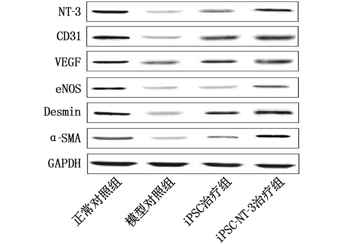
在大鼠阴茎海绵体NT-3、CD31、VEGF、eNOS、Desmin、α-SMA蛋白水平比较中,模型对照组较正常对照组降低, iPSC治疗组、iPSC-NT-3治疗组较模型对照组升高, iPSC-NT-3治疗组较iPSC治疗组升高,差异有统计学意义(P < 0.05)。见表 6、图 1。
表 6 各组大鼠治疗后阴茎海绵体NT-3、CD31、VEGF、eNOS、Desmin和α-SMA蛋白水平比较(x±s)组别 NT-3 CD31 VEGF eNOS Desmin α-SMA 正常对照组(n=8) 0.53±0.05 0.74±0.06 0.68±0.06 0.43±0.04 0.81±0.07 0.57±0.05 模型对照组(n=8) 0.16±0.03* 0.24±0.02* 0.31±0.03* 0.13±0.02* 0.34±0.03* 0.21±0.03* iPSC治疗组(n=8) 0.32±0.04# 0.37±0.05# 0.44±0.04# 0.21±0.04# 0.47±0.06# 0.32±0.04# iPSC-NT-3治疗组(n=8) 0.44±0.06#△ 0.51±0.04#△ 0.53±0.05#△ 0.35±0.04#△ 0.63±0.05#△ 0.41±0.04#△ 与正常对照组比较, *P < 0.05; 与模型对照组比较, #P < 0.05; 与iPSC治疗组比较, △P < 0.05。 3. 讨论
DIED的发病机制涉及血管内皮功能障碍、神经病变、平滑肌障碍等,但具体机制尚未完全阐明[6]。DIED发病起始于阴茎海绵体血管内皮功能障碍,并由此引发神经、平滑肌等一系列病理变化[7]。临床上ED的首选治疗方案是磷酸二酯酶抑制剂(PDE5i)类药物,其是通过减少一氧化氮合酶(NOS)/环磷酸鸟苷(cGMP)通路上cGMP的降解而发挥对ED的治疗作用。然而, DIED患者阴茎组织释放一氧化氮(NO)显著减少,但是PDE5i药物未能增加阴茎组织中NO的释放,因此约35%的DIED患者口服PDE5i治疗无效。随着干细胞在组织工程领域研究的不断深入,目前已有将脂肪源性干细胞用于DIED的研究[8-9]报道,并且PEDF转染的脂肪源性干细胞也表现出治疗勃起功能障碍的潜在价值。iPSC不仅具有自我更新与分化为三胚层细胞的能力,而且可以避免干细胞研究中遇到的伦理道德和免疫排斥等问题,在医学研究及临床应用中有着广阔的前景。NT-3是神经营养因子家族的重要成员,对神经元和功能恢复具有重要作用,可改善内源性神经营养因子不足的微环境[10-11]。
本研究结果显示, iPSC治疗组和iPSC-NT-3治疗组大鼠首次爬背时间缩短,而舔嗅次数、骑跨次数和插入次数均较模型对照组增加,并且iPSC-NT-3治疗组首次爬背时间较iPSC治疗组缩短,舔嗅次数、骑跨次数和插入次数均增加,上述差异均有统计学意义(P < 0.05), 说明iPSC和iPSC-NT-3可显著提高DIED大鼠的性功能,并且iPSC-NT-3作用优于iPSC。此外, iPSC治疗组和iPSC-NT-3治疗组大鼠ICP和ICP/MAP均较模型对照组升高, iPSC-NT-3治疗组大鼠ICP和ICP/MAP均较iPSC治疗组升高,差异均有统计学意义(P < 0.05), 说明iPSC和iPSC-NT-3可显著提高DIED大鼠的勃起功能,并且iPSC-NT-3作用优于iPSC。
氮能神经、内皮和平滑肌是阴茎正常勃起的关键。阴茎勃起是在各种刺激作用下中枢和外周神经元中NOS激活,引起阴茎海绵体内皮细胞合成和分泌NO,NO刺激cGMP的产生,并与平滑肌细胞中的cGMP结合,阴茎海绵体平滑肌细胞松弛,动脉血充满海绵体,使阴茎体积增大而勃起[12]。因此, NOS对NO的合成在阴茎勃起中具有重要作用。研究[13]发现, NOS有eNOS、神经性一氧化氮合酶(nNOS)和诱导型一氧化氮合酶(iNOS)共3种亚型,其中eNOS在勃起中起主要作用。本研究结果显示, iPSC治疗组和iPSC-NT-3治疗组大鼠阴茎海绵组织中eNOS mRNA和其蛋白表达水平均较模型对照组升高,并且iPSC-NT-3治疗组大鼠阴茎海绵组织中eNOS mRNA和其蛋白表达水平均较iPSC治疗组升高,差异有统计学意义(P < 0.05), 说明iPSC和iPSC-NT-3可显著提高DIED大鼠海绵体内eNOS的表达,并且iPSC-NT-3作用优于iPSC。因此, iPSC-NT-3可能通过提高DIED大鼠海绵体内eNOS的表达而促进海绵体NO生成,从而发挥治疗作用。
阴茎海绵体小梁间的腔隙是海绵体窦,海绵体窦的螺旋动脉直接开口于海绵体窦,在阴茎勃起中发挥重要作用。CD31在发育成熟个体的所有血管内皮细胞都有高度表达,主要位于内皮细胞之间的连接部,参与血管内皮细胞单层结构完整性的维持[14]。作为促血管生成因子, VEGF具有多种功能,包括刺激增殖、抑制凋亡和促进细胞存活, VEGF可通过促进血管内皮细胞表达而改善内皮功能,增加平滑肌数量,从而改善ED大鼠的勃起功能[15]。本研究结果显示, iPSC治疗组和iPSC-NT-3治疗组大鼠阴茎海绵组织中CD31、VEGF的mRNA及其蛋白表达水平均较模型对照组升高,并且iPSC-NT-3治疗组大鼠阴茎海绵组织中CD31、VEGF的mRNA及其蛋白表达水平均较iPSC治疗组升高,差异均有统计学意义(P < 0.05), 说明iPSC和iPSC-NT-3可显著提高DIED大鼠海绵体内CD31和VEGF的表达,并且iPSC-NT-3作用优于iPSC。因此, iPSC-NT-3可能通过提高DIED大鼠海绵体内CD31和VEGF的表达而促进海绵体血管生成,从而发挥治疗作用。
平滑肌细胞是阴茎海绵体的主要组成成分,占阴茎海绵体的40%~50%, 而阴茎内神经调控的主要效应器分布在平滑肌,性兴奋时平滑肌松弛,海绵体窦充血,阴茎变硬而勃起。本研究结果显示, iPSC治疗组和iPSC-NT-3治疗组大鼠阴茎海绵组织中Desmin、α-SMA的mRNA及其蛋白表达水平均较模型对照组升高,并且iPSC-NT-3治疗组大鼠阴茎海绵组织中Desmin、α-SMA的mRNA及其蛋白表达水平均较iPSC治疗组升高,差异均有统计学意义(P < 0.05), 说明iPSC和iPSC-NT-3可显著提高DIED大鼠海绵体内Desmin和α-SMA的表达,并且iPSC-NT-3作用优于iPSC。因此, iPSC-NT-3可能通过提高DIED大鼠海绵体内Desmin和α-SMA的表达而促进海绵体平滑肌生成,从而发挥治疗作用。
综上所述,iPSC-NT-3可通过促进海绵体组织eNOS、血管内皮及平滑肌的生成而改善DIED大鼠的性功能和勃起功能,是一种潜在的治疗DIED的方法。
-
表 1 PCR的引物序列
引物名称 引物序列 NT-3 forward: 5′- CGTGGCCATTGAACACAGCA - 3′ reverse: 5′- CCCACTGGCGGGTAAGGAAA - 3′ CD31 forward: 5′- CATGGTGGAGCACAGTGGCA - 3′ reverse: 5′- TGGGATGGAGCAGGACAGGTT - 3′ VEGF forward: 5′- CCCACCCACATACATACATT - 3′ reverse: 5′- CTCCCAACTCAAGTCCACA - 3′ eNOS forward: 5′- CTG TGC TGG CAT ACA GAA CC - 3′ reverse: 5′- CTG CCT TGA GTT GGC TCA TC - 3′ Desmin forward: 5′- CTTGATGAGGCAGATGAGGGA - 3′ reverse: 5′- AGCTTCCGGTAGGTGGCAAT - 3′ α-SMA forward: 5′- TTCCCATCCATCGTG - 3′ reverse: 5′- GCCTTAGG GTTCAGC - 3′ GAPDH forward: 5′- GAG AGG GAA ATC GTG CGT GAC-3′ reverse: 5′- CAT CTG CTG GAA GGT GGA CT-3′ 表 2 各组大鼠治疗前后血糖和体质量比较(x±s)
组别 治疗前 治疗后 体质量/g 血糖/(mmol/L) 体质量/g 血糖/(mmol/L) 正常对照组(n=8) 424.27±45.28 7.48±1.51 486.82±47.50* 7.63±1.65 模型对照组(n=8) 305.62±37.51# 28.39±4.67# 297.89±41.35# 28.79±4.56# iPSC治疗组(n=8) 298.35±41.15# 29.05±4.25# 291.46±39.71# 29.47±4.47# iPSC-NT-3治疗组(n=8) 302.40±39.67# 28.86±4.49# 295.73±42.55# 29.34±4.38# iPSC: 诱导多能干细胞; iPSC-NT-3: 神经营养素-3基因过表达的iPSC。与治疗前比较, *P < 0.05; 与正常对照组比较, #P < 0.05。 表 3 各组大鼠治疗后性功能比较(x±s)
组别 首次爬背时间/min 舔嗅次数/次 骑跨次数/次 插入次数/次 正常对照组(n=8) 42.15±4.85 39.72±5.29 15.61±2.72 9.41±1.75 模型对照组(n=8) 114.47±23.57* 13.45±2.17* 4.58±1.21* 0* iPSC治疗组(n=8) 76.21±17.24# 21.86±3.63# 6.62±1.67# 2.79±0.52# iPSC-NT-3治疗组(n=8) 51.39±15.26#△ 27.54±3.75#△ 8.95±2.12#△ 5.79±0.67#△ 与正常对照组比较, *P < 0.05; 与模型对照组比较, #P < 0.05; 与iPSC治疗组比较, △P < 0.05。 表 4 各组大鼠治疗后勃起功能的比较(x±s)
组别 ICP/mmHg ICP/MAP/% 正常对照组(n=8) 83.15±5.27 75.39±4.75 模型对照组(n=8) 47.38±3.44* 39.50±3.31* iPSC治疗组(n=8) 62.61±4.56# 52.75±4.78# iPSC-NT-3治疗组(n=8) 73.46±5.39#△ 66.32±5.17#△ ICP: 海绵窦内压; ICP/MAP: 海绵窦内压占平均动脉压的
比率。与正常对照组比较, *P < 0.05; 与模型对照组比较,
#P < 0.05; 与iPSC治疗组比较, △P < 0.05。表 5 各组大鼠治疗后阴茎海绵体NT-3、CD31、VEGF、eNOS、Desmin和α-SMA的mRNA表达水平比较(x±s)
组别 NT-3 CD31 VEGF eNOS Desmin α-SMA 正常对照组(n=8) 0.87±0.05 1.12±0.07 1.27±0.09 0.78±0.07 1.34±0.12 1.04±0.07 模型对照组(n=8) 0.25±0.03* 0.51±0.04* 0.65±0.05* 0.24±0.03* 0.67±0.05* 0.47±0.04* iPSC治疗组(n=8) 0.39±0.05# 0.65±0.05# 0.78±0.07# 0.49±0.05# 0.85±0.07# 0.69±0.05# iPSC-NT-3治疗组(n=8) 0.64±0.06#△ 0.78±0.06#△ 0.94±0.06#△ 0.62±0.04#△ 1.09±0.06#△ 0.85±0.05#△ NT-3: 神经营养素-3; VEGF: 血管内皮生长因子; eNOS: 内皮型一氧化氮合酶; Desmin: 结蛋白; α-SMA: α-平滑肌肌动蛋白。
与正常对照组比较, *P < 0.05; 与模型对照组比较, #P < 0.05; 与iPSC治疗组比较, △P < 0.05。表 6 各组大鼠治疗后阴茎海绵体NT-3、CD31、VEGF、eNOS、Desmin和α-SMA蛋白水平比较(x±s)
组别 NT-3 CD31 VEGF eNOS Desmin α-SMA 正常对照组(n=8) 0.53±0.05 0.74±0.06 0.68±0.06 0.43±0.04 0.81±0.07 0.57±0.05 模型对照组(n=8) 0.16±0.03* 0.24±0.02* 0.31±0.03* 0.13±0.02* 0.34±0.03* 0.21±0.03* iPSC治疗组(n=8) 0.32±0.04# 0.37±0.05# 0.44±0.04# 0.21±0.04# 0.47±0.06# 0.32±0.04# iPSC-NT-3治疗组(n=8) 0.44±0.06#△ 0.51±0.04#△ 0.53±0.05#△ 0.35±0.04#△ 0.63±0.05#△ 0.41±0.04#△ 与正常对照组比较, *P < 0.05; 与模型对照组比较, #P < 0.05; 与iPSC治疗组比较, △P < 0.05。 -
[1] WEN Y, LIU G H, ZHANG Y, et al. MicroRNA-205 is associated with diabetes mellitus-induced erectile dysfunction via down-regulating the androgen receptor[J]. J Cell Mol Med, 2019, 23(5): 3257-3270. doi: 10.1111/jcmm.14212
[2] DING F, SHAN C Y, LI H W, et al. Simvastatin alleviated diabetes mellitus-induced erectile dysfunction in rats by enhancing AMPK pathway-induced autophagy[J]. Andrology, 2020, 8(3): 780-792. doi: 10.1111/andr.12758
[3] 刘安全, 代苗英, 陈挺, 等. 复方玄驹胶囊联合十一酸睾酮治疗糖尿病性勃起功能障碍的疗效分析[J]. 中国性科学, 2019, 28(2): 46-48. doi: 10.3969/j.issn.1672-1993.2019.02.013 [4] 刘贵华, 张弛, 谢云, 等. 尿源干细胞治疗1型糖尿病性勃起功能障碍大鼠的实验研究[J]. 临床泌尿外科杂志, 2018, 33(8): 626-632. doi: 10.13201/j.issn.1001-1420.2018.08.009 [5] LU J, XIN Z X, ZHANG Q, et al. Beneficial effect of PEDF-transfected ADSCs on erectile dysfunction in a streptozotocin-diabetic rat model[J]. Cell Tissue Res, 2016, 366(3): 623-637. doi: 10.1007/s00441-016-2494-7
[6] SONG J Y, SUN T T, TANG Z, et al. Exosomes derived from smooth muscle cells ameliorate diabetes-induced erectile dysfunction by inhibiting fibrosis and modulating the NO/cGMP pathway[J]. J Cell Mol Med, 2020, 24(22): 13289-13302. doi: 10.1111/jcmm.15946
[7] 聂莉, 李琰华, 李俊伟. 2型糖尿病伴勃起功能障碍的研究进展[J]. 中国全科医学, 2020, 23(33): 4267-4273. doi: 10.12114/j.issn.1007-9572.2020.00.499 [8] CASTELA Â, COSTA C. Molecular mechanisms associated with diabetic endothelial-erectile dysfunction[J]. Nat Rev Urol, 2016, 13(5): 266-274. doi: 10.1038/nrurol.2016.23
[9] 燕浩, 肖冬冬, 荣立夺, 等. 脂肪来源干细胞促进糖尿病ED勃起功能恢复的实验研究[J]. 中国男科学杂志, 2019, 33(2): 24-28, 35. https://www.cnki.com.cn/Article/CJFDTOTAL-NXXX201902006.htm [10] ZHOU F, HUI Y, XIN H, et al. Therapeutic effects of adipose-derived stem cells-based microtissues on erectile dysfunction in streptozotocin-induced diabetic rats[J]. Asian J Androl, 2017, 19(1): 91-97. doi: 10.4103/1008-682X.182817
[11] 白刚, 张洪钿, 赖军, 等. NT-3-HUMSCs联合基因沉默SOCS3治疗SD大鼠脊髓损伤后的神经再生修复[J]. 昆明医科大学学报, 2018, 39(3): 31-37. doi: 10.3969/j.issn.1003-4706.2018.03.006 [12] 卢元帅, 徐宁, 杨茂光, 等. 糖尿病性勃起功能障碍发病机制研究进展[J]. 中国实验诊断学, 2019, 23(8): 1476-1478. doi: 10.3969/j.issn.1007-4287.2019.08.065 [13] 胡睿, 张威, 欧宁静, 等. 胰岛素治疗对糖尿病大鼠阴茎组织中miR-126、VEGF、eNOS表达的影响[J]. 天津医科大学学报, 2020, 26(5): 422-428. https://www.cnki.com.cn/Article/CJFDTOTAL-TJYK202005007.htm [14] PANNIER D, PHILIPPIN-LAURIDANT G, BARANZELLI M C, et al. High expression levels of egfl7 correlate with low endothelial cell activation in peritumoral vessels of human breast cancer[J]. Oncol Lett, 2016, 12(2): 1422-1428. doi: 10.3892/ol.2016.4791
[15] 提运荣, 肖冬冬, 卢慕峻. 细胞因子治疗勃起功能障碍的研究进展[J]. 中华男科学杂志, 2019, 25(7): 660-663. https://www.cnki.com.cn/Article/CJFDTOTAL-NKXB201907021.htm -
期刊类型引用(1)
1. 陆少华,杨斌斌,冯挺挺,陈碧琴,任迪龙. 神经营养因子联合他达拉非治疗2型糖尿病勃起功能障碍疗效及对血清神经生长因子脑源性神经营养因子血管内皮生长因子表达水平的影响. 中国药物与临床. 2024(10): 631-636 .  百度学术
百度学术
其他类型引用(0)


 下载:
下载:

 苏公网安备 32100302010246号