
Value of diffusion weighted imaging in preosurgical planning of pelvic malignant bone tumors
-
摘要:目的
探讨弥散加权成像(DWI)在骨盆常见恶性骨肿瘤术前规划中的应用价值。
方法回顾性分析2014年12月—2021年10月收治的经病理证实的35例骨盆软骨肉瘤和骨肉瘤患者的临床、影像和病理学资料。13例骨肉瘤患者纳入骨肉瘤组, 22例软骨肉瘤患者纳入软骨肉瘤组。通过配准CT与对应DWI图像, 借助Medraw软件进行肿瘤边界勾画,形成骨盆肿瘤三维模型报告,辅助临床设计手术截骨距离方案。记录软骨肉瘤组和骨肉瘤组患者的磁共振成像(MRI)表观弥散系数(ADC)值、术后病理切缘结果及短期影像随访复发率情况。
结果35例患者中, 26例累及髂骨, 16例累及髋臼, 8例髂骨单独受累, 15例同时累及3个或以上部位。软骨肉瘤组的平均ADC值为(1.21±0.17), 高于骨肉瘤组的(0.97±0.21), 差异有统计学意义(P < 0.000 1)。所有病例肿瘤切除骨边缘均为阴性, 6个月内临床随访均无复发。
结论DWI对骨盆恶性骨肿瘤边界判断有一定帮助,基于DWI和ADC值的术前规划截骨范围有助于设计临床个性化手术方案。
Abstract:ObjectiveTo explore the value of diffusion weighted imaging (DWI) in presurgical planning of pelvic bone neoplasms.
MethodsThe clinical, imaging and pathological data of patients pathologically confirmed as pelvic chondrosarcoma and osteosarcoma from December 2014 to October 2021 were retrospectively analyzed. Thirteen patients with osteosarcoma and 22 patients with chondrosarcoma were included in the osteosarcoma group and chondrosarcoma group. Through the registration of CT and corresponding DWI images, the tumor boundary was drawn by the Medraw software to form a three-dimensional pelvis model to design the osteotomy distance. The magnetic resonance imaging (MRI) apparent diffusion coefficient (ADC), postoperative pathological results and short-term recurrence rate of two groups were compared.
ResultsOf the 35 patients, 26 involved the ilium and 16 involved the acetabulum, of which 8 involved the ilium alone and 15 involved three or more sites simultaneously. The average ADC value in chondrosarcoma group was (1.21±0.17), which was higher than (0.97±0.21) in the osteosarcoma group (P < 0.000 1). The resection margins of the tumor were negative in all cases, and there was no recurrence within 6 months of clinical follow-up.
ConclusionDWI is helpful to judge the pelvic malignant bone tumors boundaries, and the presurgical planning of osteotomy range based on DWI and ADC value can help individualized operation design.
-
-
![]()
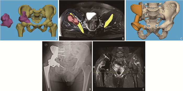
图 2 影像辅助规划手术过程
A: 骨盆肿瘤三维模型; B: 基于CT和MRI配准的融合重建肿瘤模型(图中蓝线为术前模拟10、20、25、30 mm截骨范围); C: 假体和骨盆模型; D: 术后骨盆平片; E: 术后6个月的MRI图。
表 1 2组患者基本临床信息比较(x±s)[n(%)]
临床特征 骨肉瘤组(n=13) 软骨肉瘤组(n=22) 年龄/岁 45.08±18.63 51.64±13.26 男 8(61.54) 17(77.27) 女 5(38.46) 5(22.73) 肿瘤最大直径/cm 13.85±5.01 13.75±5.81 ADC值/(×10-3 mm2/s) 0.97±0.21* 1.21±0.17 术后随访原位复发 0 0 ADC: MRI表观弥散系数。与软骨肉瘤组比较, * P<0.000 1。  下载: 导出CSV
-
[1] LEE J S, KELLY C M, BARTLETT E K. Management of pelvic sarcoma[J]. Eur J Surg Oncol, 2022, 48(11): 2299-2307. doi: 10.1016/j.ejso.2022.09.011
[2] HU X, LU M, WANG J, et al. Combined and modified Gibson and ilioinguinal approaches in type Ⅱ+Ⅲ internal hemipelvectomy for periacetabular tumors[J]. Front Oncol, 2022, 12: 934812. doi: 10.3389/fonc.2022.934812
[3] XU L, QIN H, TAN J, et al. Clinical study of 3D printed personalized prosthesis in the treatment of bone defect after pelvic tumor resection[J]. J Orthop Translat, 2021, 29: 163-169. doi: 10.1016/j.jot.2021.05.007
[4] QU Y, LI X, YAN Z, et al. Surgical planning of pelvic tumor using multi-view CNN with relation-context representation learning[J]. Med Image Anal, 2021, 69: 101954. doi: 10.1016/j.media.2020.101954
[5] FUJIWARA T, MEDELLIN RINCON M R, SAMBRI A, et al. Limb-salvage reconstruction following resection of pelvic bone sarcomas involving the acetabulum[J]. Bone Joint J, 2021, 103-B(4): 795-803. doi: 10.1302/0301-620X.103B4.BJJ-2020-0665.R1
[6] 刘广辉. 磁共振弥散加权成像评估骨肌肿瘤的临床分析[J]. 实用医学影像杂志, 2022, 23(1): 84-87. https://www.cnki.com.cn/Article/CJFDTOTAL-SYXY202201029.htm [7] CHHABRA A, ASHIKYAN O, SLEPICKA C, et al. Conventional MR and diffusion-weighted imaging of musculoskeletal soft tissue malignancy: correlation with histologic grading[J]. Eur Radiol, 2019, 29(8): 4485-4494. doi: 10.1007/s00330-018-5845-9
[8] 李莹, 任翠萍, 程敬亮, 等. 磁共振动态增强及扩散加权成像对骨肉瘤恶性程度评判的价值[J]. 临床放射学杂志, 2018, 37(4): 666-669. https://www.cnki.com.cn/Article/CJFDTOTAL-LCFS201804029.htm [9] KANG Y, YUAN W, DING X, et al. Chondrosarcoma of the para-acetabulum: correlation of imaging features with histopathological grade[J]. La Radiologia Medica, 2016, 121(12): 897-904. doi: 10.1007/s11547-016-0673-y
[10] 曲扬, 艾松涛, 杨飞, 等. CT和MRI图像配准融合联合3D打印技术在难治性骨盆肿瘤术前规划中的应用[J]. 上海交通大学学报: 医学版, 2017, 37(9): 1239-1244, 1248. https://www.cnki.com.cn/Article/CJFDTOTAL-SHEY201709011.htm [11] GILL J, GORLICK R. Advancing therapy for osteosarcoma[J]. Nat Rev Clin Oncol, 2021, 18(10): 609-624. doi: 10.1038/s41571-021-00519-8
[12] WANG J, MIN L, LU M, et al. What are the Complications of Three-dimensionally Printed, Custom-made, Integrative Hemipelvic Endoprostheses in Patients with Primary Malignancies Involving the Acetabulum, and What is the Function of These Patients[J]. Clin Orthop Relat Res, 2020, 478(11): 2487-2501. doi: 10.1097/CORR.0000000000001297
[13] JI T, YANG Y, TANG X, et al. 3D-Printed Modular Hemipelvic Endoprosthetic Reconstruction Following Periacetabular Tumor Resection: Early Results of 80 Consecutive Cases[J]. J Bone Joint Surg Am, 2020, 102(17): 1530-1541. doi: 10.2106/JBJS.19.01437
[14] ASMAR K, SAADE C, SALMAN R, et al. The value of diffusion weighted imaging and apparent diffusion coefficient in primary Osteogenic and Ewing sarcomas for the monitoring of response to treatment: initial experience[J]. Eur J Radiol, 2020, 124: 108855. doi: 10.1016/j.ejrad.2020.108855
[15] HONG J H, JEE W H, JUNG C K, et al. Soft tissue sarcoma: adding diffusion-weighted imaging improves MR imaging evaluation of tumor margin infiltration[J]. Eur Radiol, 2019, 29(5): 2589-2597. doi: 10.1007/s00330-018-5817-0
[16] YUAN Y, ZENG D, LIU Y, et al. DWI and IVIM are predictors of Ki67 proliferation index: direct comparison of MRI images and pathological slices in a murine model of rhabdomyosarcoma[J]. Eur Radiol, 2020, 30(3): 1334-1341. doi: 10.1007/s00330-019-06509-w
[17] 潘献伟, 刘泳坚. 原发性骨肉瘤影像学表现与病理分型的关系[J]. 临床骨科杂志, 2021, 24(1): 51-54. https://www.cnki.com.cn/Article/CJFDTOTAL-LCGK202101021.htm [18] 柳思宇, 吴兵, 李小敏, 等. 弥散加权成像对隆突性皮肤纤维肉瘤术前规划的价值初探[J]. 上海交通大学学报: 医学版, 2022, 42(8): 1095-1102. https://www.cnki.com.cn/Article/CJFDTOTAL-SHEY202208014.htm [19] YOON M A, CHEE C G, CHUNG H W, et al. Added value of diffusion-weighted imaging to conventional MRI for predicting fascial involvement of soft tissue sarcomas[J]. Eur Radiol, 2019, 29(4): 1863-1873. doi: 10.1007/s00330-018-5786-3
-
期刊类型引用(9)
1. 马雪萍,王晓丽,阿里木江·司马义,徐桂萍. 腹股沟上髂筋膜阻滞复合全身麻醉对高龄髋关节置换患者术后谵妄发生的影响. 新疆医学. 2022(02): 131-134 .  百度学术
百度学术
2. 张宏,李淑萍. 老年患者髋关节置换术后谵妄的发生现状及其相关影响因素分析. 长春中医药大学学报. 2022(10): 1155-1159 . 百度学术
3. 刘贵政,郑婷婷,杜斌. 老年髋部骨折患者术后谵妄发生现况及危险因素研究. 贵州医药. 2022(09): 1405-1406 . 百度学术
4. 王秀环,鲍乐乐,马漪洁,陈宁宁. 不同麻醉方法对老年髋关节置换患者术后谵妄发生的影响. 广州医科大学学报. 2021(02): 40-44 . 百度学术
5. 刘丹,杨万翔. 人文关怀护理对人工髋关节置换术后谵妄患者临床症状的影响. 中国当代医药. 2021(25): 270-272+276 . 百度学术
6. 陈立红,徐芙蓉,叶洁玉,许华亮. 高龄骨科髋关节置换术后患者发生谵妄的危险因素分析. 现代医学与健康研究电子杂志. 2021(23): 115-118 . 百度学术
7. 毛俊岚. 高龄患者髋关节置换术后谵妄1例的护理. 基层医学论坛. 2020(03): 420-421 . 百度学术
8. 欧玉琼,吴建颖,周嫦娥. 以临床路径为指导的谵妄管理对老年股骨头置换术后患者的影响. 护理实践与研究. 2020(02): 86-88 . 百度学术
9. 姜红卫. 骨科老年患者髋关节术后谵妄发生原因及疼痛干预护理进展. 系统医学. 2020(13): 196-198 . 百度学术
其他类型引用(1)
计量
- 文章访问数: 208
- HTML全文浏览量: 47
- PDF下载量: 25
- 被引次数: 10
 苏公网安备 32100302010246号