Value of artificial intelligence combined with super microvascular imaging technology in the diagnosis of breast nodules
-
摘要:目的
探讨人工智能S-Detect技术联合智能三维超微血管成像(3D-SMI)技术对乳腺结节良恶性的诊断价值。
方法选取2021年1月—2023年2月151例(192个结节)乳腺病变患者为研究对象。采用常规超声检查、S-Detect技术、智能3D-SMI技术对乳腺结节进行良恶性鉴别, 以术后病理结果为金标准,绘制受试者工作特征(ROC)曲线,分析常规超声检查、S-Detect技术、智能3D-SMI技术及三者联合诊断对乳腺结节良恶性的诊断效能。
结果192个结节中,病理证实良性结节112个,恶性结节80个。常规超声检查、S-Detect技术、智能3D-SMI技术及三者联合诊断的敏感度、特异度和准确度分别为70.00%、83.93%、78.13%, 78.75%、79.46%、79.17%, 71.25%、93.75%、84.38%, 90.00%、80.36%、84.38%。三者联合诊断的诊断效能较常规超声检查、S-Detect技术高,差异有统计学意义(Z=2.567, P=0.010; Z=2.533, P=0.011)。常规超声检查、S-Detect技术、智能3D-SMI技术的曲线下面积(AUC)比较,差异无统计学意义(P>0.05)。
结论人工智能S-Detect技术与智能3D-SMI技术联合应用可辅助常规超声诊断乳腺结节的良恶性,有助于提高诊断的准确率。
-
关键词:
- S-Detect技术 /
- 超微血管成像 /
- 乳腺肿瘤 /
- 超声检查
Abstract:ObjectiveTo explore the diagnostic value of artificial intelligence S-Detect technique and smart three-dimensional super microvascular imaging (3D-SMI) technique for diagnosis of benign and malignant breast nodules.
MethodsA total of 151 patients with breast lesions (192 nodules) in our hospital from January 2021 to February 2023 were selected as study objects. Conventional ultrasound examination, S-Detect technique, smart 3D-SMI technique were used to identify benign and malignant breast nodules, with the postoperative pathological results as the gold standard, the receiver operating characteristic(ROC) curve was drawn, the diagnostic efficacy of conventional ultrasonography, S-Detect technology, smart 3D-SMI technique and the combined diagnosis of three techniques in the differentiation of benign and malignant breast nodules was compared.
ResultsOf 192 nodules, 112 nodules were diagnosed as benign ones and 80 as malignant ones. The sensitivity, specificity and accuracy of conventional ultrasound examination, S-Detect technique, smart 3D-SMI technique and the combination were 70.00%, 83.93%, 78.13%; 78.75%, 79.46%, 79.17%; 71.25%, 93.75% and 84.38%; 90%, 80.36% and 84.38%, respectively. The diagnostic efficacy of the their combined diagnosis was higher than that of conventional ultrasound examination and S-Detect technology (Z=2.567, P=0.010; Z=2.533, P=0.011). There was no significant difference in area under the curve(AUC) among conventional ultrasound examination, S-Detect technique and smart 3D-SMI technique (P>0.05).
ConclusionThe combined application of artificial intelligence S-Detect technique and smart 3D-SMI technique can assist the conventional ultrasound in the diagnosis of benign and malignant breast nodules, and help to improve the diagnostic accuracy.
-
Keywords:
- S-Detect technique /
- super microvascular imaging /
- breast tumor /
- ultrasonography
-
乳腺癌是中国女性最常见的肿瘤,发病率位居女性恶性肿瘤首位,严重影响患者身心健康[1]。早发现、早诊断乳腺癌有助于患者进行早期有效治疗,改善预后[2]。常规超声检查作为乳腺癌早期筛查的主要影像检查,其成本低、无辐射、分辨率高,能够使患者尽早发现肿块,实现采用更保守的手段进行治疗[3]。但常规超声诊断乳腺影像学报告和数据系统(BI-RADS)受医师经验等主观性因素影响,具有一定局限性[4]。S-Detect技术是一种基于深度学习的人工智能影像辅助诊断技术,一定程度避免了医师常规超声检查主观性强的误诊风险[5]。研究[6-7]表明,乳腺结节微血流评估有助于肿瘤良恶性的鉴别诊断,智能3D-SMI技术通过三维图像重建显示病变血流架构,提供更立体全面的微血流信息,有助于鉴别乳腺结节的良恶性。本研究旨在评估智能3D-SMI技术与S-Detect技术联合应用辅助常规超声检查诊断的应用价值。
1. 资料与方法
1.1 一般资料
选取2021年1月—2023年2月上海交通大学医学院附属第九人民医院黄浦分院的151例女性患者为研究对象,共192个结节,直径0.40~4.50 cm, 平均(1.85±0.90) cm; 患者年龄19~80岁,平均(52.72±14.03)岁。纳入标准: ①所有患者均经术后病理证实,术前均由1名10年以上工作经验的医师行常规超声、S-Detect技术和智能3D-SMI技术检查并完整留图; ②肿块最大直径 < 5 cm者。排除既往有手术史、放化疗史以及怀孕和哺乳期患者。本研究经医院伦理委员会批准。
1.2 仪器与方法
1.2.1 常规超声、3D-SMI技术检查
应用Toshiba Aplio 900超声仪(L14-5探头,频率5~14 MHz)进行检查。患者取仰卧位,充分暴露乳房,并对每个象限进行多层面扫描。1名具有10年以上工作经验的超声医生进行检查、留图,测量病变部位的最大直径,并记录病变的大小、形态、方向、内部回声、钙化等重要的超声特征并存图3D-SMI血流架构。在双盲法下,由2名超过10年丰富工作经验的超声医生独立对每个病变根据病灶二维超声特征进行BI-RADS分类,记录结果并对3D-SMI血流架构图进行分类。诊断不一致时,咨询主任医师,并最终协商达成统一诊断。
1.2.2 S-Detect技术检查:
使用三星RS80A型超声仪(LA523探头,频率5~13 MHz)进行检测。选取最大长径切面及横断面分别冻结,并启动内置的S-Detect模式,病变可被诊断为良性或恶性。当最大长径和横断面诊断不一致时,分为2种诊断方式: 方式1(只要1个诊断结果为良性,则S-Detect技术诊断结果为良性)及方式2(只要1个诊断结果为恶性,则S-Detect技术诊断结果为恶性)。比较2种方式的诊断效能,选取诊断效能高的诊断方式作为S-Detect技术鉴别结节良恶性的最终诊断方式。
1.2.3 智能3D-SMI血流架构的评估
基于智能3D-SMI与其他乳腺微血管形态的研究,将血流架构分为5型[8], Ⅰ型(无血管)、Ⅱ型(血流架构呈线状,病变内见无交叉的少量血管)、Ⅲ型(血流架构呈树枝状,可见病变内微血管有分支)、Ⅳ型(血流架构呈残根状,微血管排列扭曲,病变周围粗大扭曲的血管 < 2根)、Ⅴ型(血流架构呈蟹足状,病变周围粗大扭曲的血管≥2根)。本研究将血流架构分型为Ⅰ型、Ⅱ型、Ⅲ型的结节判断为良性结节,将血流架构分型为Ⅳ型和Ⅴ型的结节判断为恶性结节。
1.2.4 三者联合诊断
若S-Detect技术诊断为良性, 3D-SMI技术诊断为恶性,则联合诊断结果为恶性; 若3D-SMI技术诊断为良性, S-Detect技术诊断为恶性,则将BI-RADS分类升高1级,本实验设定BI-RADS分类≤4 a的结节为良性, ≥4 b的结节为恶性。
1.3 统计学方法
采用SPSS 22.0软件进行统计学分析,采用卡方检验分别比较不同组别的诊断效能,以病理结果为金标准,绘制受试者工作特征(ROC)曲线,计算每条曲线的曲线下面积(AUC), 应用Z值评估各组间AUC的差异, P < 0.05为差异有统计学意义。
2. 结果
2.1 病理结果
192个结节中,恶性80个(浸润性导管癌63个,导管原位癌4个,导管内癌伴微小浸润3个,乳头状癌7个,浸润性小叶癌2个,黏液癌1个)和良性112个(纤维腺瘤66个,硬化性乳腺病3个,导管内乳头状瘤14个,乳腺增生2个,间质胶原化9个,乳腺囊肿伴炎症4个,错构瘤2个,管状腺瘤1个,肌上皮瘤1个,良性叶状肿瘤1个,炎症5个,脂肪坏死4个)。
2.2 S-Detect技术不同诊断方式的结果
S-Detect技术诊断方式1和方式2的AUC值分别为0.791、0.741, 方式1的诊断效能较方式2高,差异有统计学意义(Z=2.048, P=0.041), 因此选择方式1作为S-Detect技术的最终诊断方式。
2.3 常规超声检查、S-Detect技术、智能3D-SMI技术及三者联合检查结果
常规超声检查诊断的良性结节118个,恶性结节74个,敏感度、特异度和准确度分别为70.00%、83.93%、78.13%; S-Detect技术检查诊断的良性结节106个,恶性结节86个,敏感度、特异度和准确度分别为78.75%、79.46%、79.17%; 智能3D-SMI技术诊断的良性结节128个,恶性结节64个,敏感度、特异度和准确度分别为71.25%、93.75%、84.38%; 三者联合诊断的良性结节98个,恶性结节94个,敏感度、特异度和准确度分别为90.00%、80.36%、84.38%。见表 1和表 2。
表 1 不同检查方法诊断良恶性结节的结果例 方法 结果 病理结果 合计 恶性(n=80) 良性(n=112) 常规超声检查 恶性 56 18 74 良性 24 94 118 S-Detect技术 恶性 63 23 86 良性 17 89 106 智能3D-SMI技术 恶性 57 7 64 良性 23 105 128 三者联合诊断 恶性 72 22 94 良性 8 90 98 表 2 不同检查方法对乳腺良恶性结节的诊断效能方法 敏感度/% 特异度/% 准确度/% 阳性预测值/% 阴性预测值/% 常规超声检查 70.00 83.93 78.13 75.68 79.66 S-Detect技术 78.75 79.46 79.17 73.26 83.96 智能3D-SMI技术 71.25 93.75 84.38 89.06 82.03 三者联合诊断 90.00 80.36 84.38 76.60 91.84 2.4 不同方法的ROC曲线和AUC值
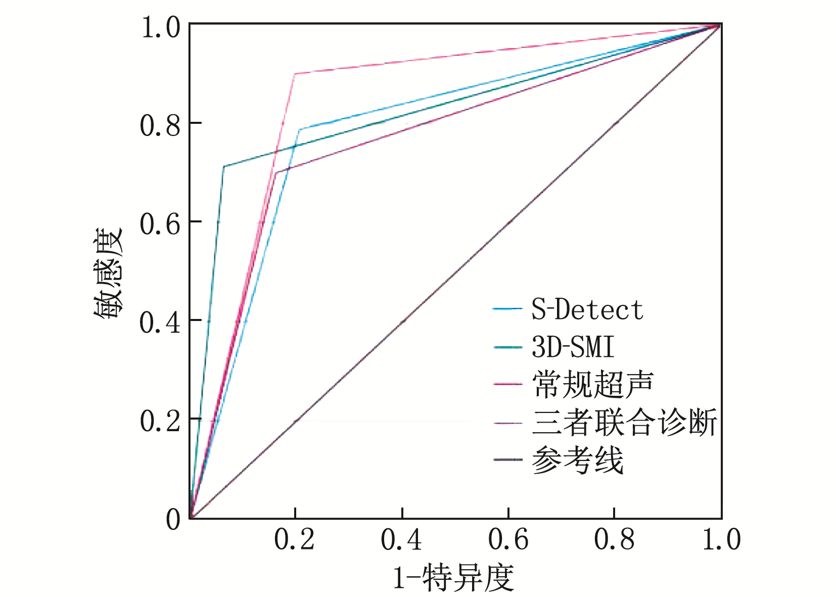
三者联合诊断、常规超声检查、S-Detect技术和智能3D-SMI技术的AUC值分别为0.852、0.770、0.791、0.825, 三者联合诊断的诊断效能较常规超声、S-Detect技术高,差异有统计学意义(Z=2.567, P=0.010; Z=2.533, P=0.011); 三者联合诊断较智能3D-SMI技术的诊断效能高,但差异无统计学意义(Z=-0.982, P=0.326), 其他组间诊断效能差异无统计学意义(P>0.05), 见图 1。
3. 讨论
乳腺癌作为女性最常见的恶性肿瘤,发病率逐年上升,对患者身心健康产生严重影响,迫切需要全面的影像检查方法在早期准确鉴别乳腺结节的良恶性[9]。常规超声检查在乳腺结节的良恶性鉴别中扮演着重要角色,但良恶性结节的部分影像特点具有重合性,且容易受医师主观性的影响,具有一定的局限性[10]。本研究首次将智能3D-SMI技术与S-Detect技术联合应用辅助常规超声鉴别乳腺结节的良恶性,避免了单一技术检查的缺陷和不足,联合应用明显提高了乳腺结节良恶性鉴别的诊断效能,目前国内尚未报道。
S-Detect计算机辅助算法基于人工智能和神经网络,对大量的训练数据进行“学习”,根据BI-RADS词汇分析乳腺结节的形态学特征。最终提供了二分法的评估: “可能良性”或“可能恶性”[11]。一些研究[12-14]已经证实, S-Detect技术提高了对乳腺结节,甚至小结节(< 2 cm)良恶性诊断的准确性。本研究151例患者的192个结节中, 80个结节经病理检查证实为恶性, 112个结节经病理检查证实为良性。S-Detect技术提示恶性结节86个,良性结节106个,其中40个被误诊。S-Detec技术诊断乳腺结节良恶性的敏感度、特异度和准确度分别为78.75%、79.46%、79.17%。17个误诊为良性的恶性结节中, 8个结节直径 < 1 cm, 边界尚清, 2个结节内微钙化,误诊的原因可能与体积太小、恶性征象不明显而无法被自动分析有关,而S-Detect技术的已知局限性是其不能分析结节内的微钙化、血流等参数。一些被S-Detect技术所忽视的微钙化部分可以被超声医生从二维图像中观察出来。误诊为恶性的良性结节23个: 纤维腺瘤13个,导管内乳头状瘤4个,乳腺炎3个,间质胶原化1个,肌上皮瘤1个,硬化性腺病1个。造成诊断不准确的原因可能与肿块的位置和深度有关[15]。部分良性结节形状不规则、边缘不光整、边缘成角或毛刺导致S-Detect程序错误定义结节的边界,可能歪曲结节的边缘,导致误诊为恶性。本研究发现, S-Detect技术的诊断效能未较常规超声检查增高,分析原因可能为样本量较少,且S-Detect技术受限于超声医师主观图像切面的选择。
SMI是一项通过自适应算法进行滤波处理杂波信号的新技术,可以显示CDFI图像中难以显示的低速血流,智能3D-SMI通过2D-SMI重建,更加直观地评估微血流架构,较2D-SMI显示更多的微血管及更全面、立体的血流信号[16]。恶性病变因细胞缺氧、增殖较快,诱导大量粗大、扭曲的新生血管生成,因此血流架构Ⅳ型和Ⅴ型为其特异性血流征象,良性病变较恶性病变生长缓慢,出现血管畸形和动静脉瘘的概率较小,因此血流架构Ⅰ型、Ⅱ型和Ⅲ型为良性病变特异性血流征象,并且具有较好的观察者一致性,重复性高[17], 使其通过血流架构判断乳腺结节良恶性成为可能。本研究智能3D-SMI技术提示恶性结节64个,良性结节128个,30个被误诊,原因可能与病灶直径及位置深浅有关,这是影响血流信号显示的重要因素。随着肿块接近皮肤表面及最大直径的增大,智能3D-SMI技术的良恶性诊断效能逐渐降低[18]。部分假阳性病变为血流丰富的炎症,被误诊为恶性,部分假阴性病变为乏血供类肿瘤易被误诊为恶性,如黏液癌,这些都会导致一定误诊率,因此需要多种检查技术相互结合提高诊断的准确性。
虽然本研究认为, S-Detect技术对乳腺良恶性病变的鉴别诊断具有明确临床价值,但仍有一定局限性。具体来说, S-Detect技术不能识别病变内的微钙化,但微钙化是恶性肿瘤可疑的重要指标,而且该技术不能提示病变的血流情况, S-Detect技术可以自动识别结节,但如果结节很小或边界不清,可能导致不适当的分类; 添加血流信息、弹性成像、萤火虫技术等其他参数可以进一步提高诊断的准确性[19]。本研究采用3种技术联合诊断,减少了单一技术检查的局限性,提供了结节的空间血流形态,提高了良恶性结节鉴别的诊断效能。本研究的不足在于样本量较少,应当扩大样本量,进行多中心研究。还应该针对分子亚型,运用S-Detect技术与智能3D-SMI技术从分子生物学角度更全面分析、研究其诊断效能。人工智能S-Detect技术与智能3D-SMI技术联合应用对乳腺结节良恶性有较高的诊断效能,值得在临床中推广。
-
表 1 不同检查方法诊断良恶性结节的结果
例 方法 结果 病理结果 合计 恶性(n=80) 良性(n=112) 常规超声检查 恶性 56 18 74 良性 24 94 118 S-Detect技术 恶性 63 23 86 良性 17 89 106 智能3D-SMI技术 恶性 57 7 64 良性 23 105 128 三者联合诊断 恶性 72 22 94 良性 8 90 98 表 2 不同检查方法对乳腺良恶性结节的诊断效能
方法 敏感度/% 特异度/% 准确度/% 阳性预测值/% 阴性预测值/% 常规超声检查 70.00 83.93 78.13 75.68 79.66 S-Detect技术 78.75 79.46 79.17 73.26 83.96 智能3D-SMI技术 71.25 93.75 84.38 89.06 82.03 三者联合诊断 90.00 80.36 84.38 76.60 91.84 -
[1] 何思怡, 李贺, 曹毛毛, 等. 全球及我国女性乳腺癌疾病负担年龄分布及变化趋势[J]. 中国肿瘤, 2023, 32(1): 1-7. https://www.cnki.com.cn/Article/CJFDTOTAL-ZHLU202301001.htm [2] ALAREF A, HASSAN A, SHARMA KANDEL R, et al. Magnetic resonance imaging features in different types of invasive breast cancer: a systematic review of the literature[J]. Cureus, 2021, 13(3): e13854.
[3] LIU M J, MEN Y M, ZHANG Y L, et al. Improvement of diagnostic efficiency in distinguishing the benign and malignant thyroid nodules via conventional ultrasound combined with ultrasound contrast and elastography[J]. Oncol Lett, 2017, 14(1): 867-871. doi: 10.3892/ol.2017.6245
[4] STROBEL K, SCHRADING S, HANSEN N L, et al. Assessment of BI-RADS category 4 lesions detected with screening mammography and screening US: utility of MR imaging[J]. Radiology, 2015, 274(2): 343-351. doi: 10.1148/radiol.14140645
[5] 戚瑞祥, 朱罗茜, 方建华, 等. S-Detect技术在乳腺非肿块型病变中的诊断价值[J]. 中国超声医学杂志, 2021, 37(3): 251-255. doi: 10.3969/j.issn.1002-0101.2021.03.004 [6] ZHANG X Y, ZHANG L, LI N, et al. Vascular index measured by smart 3-D superb microvascular imaging can help to differentiate malignant and benign breast lesion[J]. Cancer Manag Res, 2019, 11: 5481-5487. doi: 10.2147/CMAR.S203376
[7] 张曼琪, 查海玲, 刘心培, 等. 超声检查评估乳腺癌新辅助化疗疗效的准确性及影响因素分析[J]. 实用临床医药杂志, 2022, 26(13): 45-49, 54. doi: 10.7619/jcmp.20220387 [8] 薛雯, 杨柳茵, 范丽, 等. 超微血管成像技术鉴别乳腺良恶性病变[J]. 中国医学影像技术, 2019, 35(1): 77-81. https://www.cnki.com.cn/Article/CJFDTOTAL-ZYXX201901026.htm [9] GUO R R, LU G L, QIN B J, et al. Ultrasound imaging technologies for breast cancer detection and management: a review[J]. Ultrasound Med Biol, 2018, 44(1): 37-70. doi: 10.1016/j.ultrasmedbio.2017.09.012
[10] LY A, ONO J C, HUGHES K S, et al. Fine-needle aspiration biopsy of palpable breast masses: patterns of clinical use and patient experience[J]. J Natl Compr Canc Netw, 2016, 14(5): 527-536. doi: 10.6004/jnccn.2016.0061
[11] BARTOLOTTA T V, ORLANDO A A M, SPATAFORA L, et al. S-Detect characterization of focal breast lesions according to the US BI RADS lexicon: a pictorial essay[J]. J Ultrasound, 2020, 23(2): 207-215. doi: 10.1007/s40477-020-00447-w
[12] 邢博缘, 董梦, 刘冬婷, 等. 超声S-Detect技术对直径≤2 cm乳腺小结节的诊断价值[J]. 中国超声医学杂志, 2022, 38(6): 626-629. doi: 10.3969/j.issn.1002-0101.2022.06.009 [13] ZHAO C Y, XIAO M S, LIU H, et al. Reducing the number of unnecessary biopsies of US-BI-RADS 4a lesions through a deep learning method for residents-in-training: a cross-sectional study[J]. BMJ Open, 2020, 10(6): e035757. doi: 10.1136/bmjopen-2019-035757
[14] 曹力, 张会萍, 周毓青, 等. 不同年资超声科医师应用S-Detect技术诊断乳腺结节的价值研究[J]. 肿瘤影像学, 2022, 31(4): 439-443. https://www.cnki.com.cn/Article/CJFDTOTAL-YXYX202204013.htm [15] FUJIOKA T, KUBOTA K, MORI M, et al. Distinction between benign and malignant breast masses at breast ultrasound using deep learning method with convolutional neural network[J]. Jpn J Radiol, 2019, 37(6): 466-472. doi: 10.1007/s11604-019-00831-5
[16] 李伟伟, 周庆华, 吴迎, 等. 超微血管三维立体成像技术对乳腺肿块血流的评估价值[J]. 中国超声医学杂志, 2019, 35(7): 587-589. doi: 10.3969/j.issn.1002-0101.2019.07.005 [17] 蔡思曼, 王红燕, 张晓燕, 等. 智能三维超微血管成像评估乳腺病变血流的观察者间一致性分析[J]. 中华超声影像学杂志, 2020, 29(7): 613-617. [18] 肖露, 褚雯, 王华. 超微血管成像技术对乳腺肿瘤血管形态分布特征及其诊断效能的初步分析[J]. 中华超声影像学杂志, 2018, 27(11): 973-976. https://cdmd.cnki.com.cn/Article/CDMD-10464-1019658790.htm [19] CHOI J S, HAN B K, KO E S, et al. Effect of a deep learning framework-based computer-aided diagnosis system on the diagnostic performance of radiologists in differentiating between malignant and benign masses on breast ultrasonography[J]. Korean J Radiol, 2019, 20(5): 749-758.
-
期刊类型引用(7)
1. 陈凡杰,胡秀红,王萍. 超微血管成像联合剪切波弹性成像在乳腺影像学报告及数据系统4类结节诊断中的应用价值. 大医生. 2025(05): 123-125 .  百度学术
百度学术
2. 康林,刘喜,程浩,周金池,马鹏,窦维佳,刘震雄. 人工智能在早期食管癌内镜诊断中的应用进展. 空军军医大学学报. 2024(04): 465-471 .  百度学术
百度学术
3. 袁杰,汪成,笪应芬,吴林生. 联合应用二维、三维超微血管成像修正S-Detect分类良、恶性乳腺囊实性病变. 中国医学影像技术. 2024(04): 545-548 .  百度学术
百度学术
4. 袁杰,汪成,笪应芬. 萤火虫成像联合应变弹性成像校正人工智能S-Detect技术对乳腺囊实性肿块良恶性的诊断价值. 中国医疗器械杂志. 2024(04): 426-429+439 .  百度学术
百度学术
5. 丛小宇,笪应芬,胡佳飞,汪成. S-Detect技术辅助校正BI-RADS分类鉴别诊断乳腺黏液癌. 中华实用诊断与治疗杂志. 2024(09): 871-876 .  百度学术
百度学术
6. 钟树兴,郭红梅,刘美玲,王霞. 多模态超声联合人工智能S-Detect技术对BI-RADS 4类乳腺结节的诊断分析. 影像技术. 2024(05): 4-9+25 .  百度学术
百度学术
7. 叶绮婷,吴斯琪,孙秋水,张彩华,黄小玲,梁劲松. 人工智能S-Detect技术辅助诊断乳腺良恶性结节的应用价值. 影像研究与医学应用. 2023(24): 67-70 .  百度学术
百度学术
其他类型引用(1)


 下载:
下载:

 苏公网安备 32100302010246号