Diagnostic value of dynamic contrast-enhanced magnetic resonance imaging and signal intensity-time curve in benign and malignant prostate lesions
-
摘要:目的
探讨磁共振动态增强扫描(DCE-MRI)及信号强度-时间曲线(SI-T曲线)对前列腺良恶性病变的诊断价值。
方法选取102例前列腺疾病患者为研究对象,并分为良性组65例和恶性组37例。采用受试者工作特征(ROC)曲线分析DCE-MRI、SI-T检测及二者联合检测对前列腺良恶性病变的诊断价值。
结果恶性组患者的容量转移常数(Ktrans)、速率常数(Kep)、血管外细胞外间隙容积比(Ve)大于良性组,差异有统计学意义(P < 0.05)。恶性组患者的SI-T曲线速升缓降型占比高于良性组,持续缓升型、缓升平台型占比低于良性组,差异有统计学意义(P < 0.05)。恶性组患者的达峰时间短于良性组,强化程度、强化率高于良性组,差异有统计学意义(P < 0.05);DCE-MRI指标及SI-T曲线参数联合检测诊断前列腺良恶性病变的曲线下面积(AUC)大于DCE-MRI指标、SI-T曲线参数单独检测,差异有统计学意义(P < 0.05)。
结论DCE-MRI、SI-T曲线参数联合检测在前列腺良恶性病变中具有较好的诊断价值。
Abstract:ObjectiveTo explore the diagnostic value of dynamic contrast-enhanced magnetic resonance imaging (DCE-MRI) and signal intensity-time (SI-T) curve in benign and malignant prostate lesions.
MethodsA total of 102 patients with prostate disease were selected as the study objects, and were divided into benign group (65 cases) and malignant group (37 cases). Receiver operating characteristic (ROC) curve was used to analyze the diagnostic value of DCE-MRI, SI-T alone and their combined detection in benign and malignant prostate lesions.
ResultsThe volume transfer constant (Ktrans), rate constant (Kep) and extravascular extracellular space volume fraction (Ve) of patients in the malignant group were higher than those in the benign group (P < 0.05). The proportion of fast growth and slow decline of SI-T curve in the malignant group was significantly higher than that in the benign group, and the proportion of continuous slow rise and slow rise platform type was significantly lower than that in the benign group (P < 0.05). The peak time of the malignant group was significantly shorter than that of the benign group, and the degree and rate of reinforcement were significantly higher than those of the benign group (P < 0.05). The area under the curve (AUC) of the combined detection of DCE-MRI or SI-T curve parameter in the diagnosis of benign and malignant prostate lesions was greater than that of the single detection of DCE-MRI or SI-T curve parameter (P < 0.05).
ConclusionThe combined detection of DCE-MRI and SI-T curve has good diagnostic value in diagnosis of benign and malignant prostate lesions.
-
前列腺是男性生殖系统中的附属腺体,具有分泌激素及控制排尿等功能。目前,中国前列腺恶性病变的发病率及致死率呈连续增长趋势。前列腺良性病变可直接影响患者排尿功能,前列腺癌可加重患者排尿功能异常程度, 2种疾病症状相似,难以通过常规MRI进行诊断[1]。磁共振动态增强扫描(DCE-MRI)是基于MRI发展而来的一种检查技术,在注射对比剂后,通过观察对比剂在血管、组织中的分布及清除情况,以评价病变性质[2]。信号强度-时间曲线(SI-T曲线)可反映DCE-MRI信号强度随时间变化情况,能量化评价病变组织血供特点。相关研究[3]指出,前列腺癌存在DCE-MRI早期、快速及明显强化。前列腺癌组织微血管丰富,且微血管系统通透性高,新生毛细血管具有不连续的基底膜,可使对比剂进入肿瘤组织。基于此,本研究探讨DCE-MRI及SI-T曲线诊断前列腺良恶性病变的价值,以期为该疾病的临床早期诊断提供参考依据。
1. 资料与方法
1.1 一般资料
选取2020年3月—2022年5月前列腺疾病患者102例为研究对象,并分为良性组65例和恶性组37例。良性组: 年龄41~69岁,平均(56.08±5.14)岁。恶性组: 年龄37~68岁,平均(55.85±5.27)岁。2组年龄比较,差异无统计学意义(P>0.05)。
纳入标准: ①符合有关前列腺增生、前列腺癌等诊断标准[4], 存在尿频、尿急等排尿异常相关症状者; ②临床资料完整者; ③行手术治疗者。排除标准: ①存在泌尿系统畸形者; ②急性前列腺炎患者; ③既往具有腹部手术史患者; ④检查前已进行放化疗治疗者。
1.2 方法
采用飞利浦Achieva 3.0T超导MRI扫描仪及腹部相控阵线圈进行腹部检查,患者取仰卧位,膀胱适当充盈,对前列腺及精囊腺进行扫描,先行常规序列扫描[T1WI横轴位、T2WI横轴位、T2WI矢状位,层厚4.0 mm, 层间距4.5 mm, 视野(FOV): 250 mm×250 mm], 然后行DCE-MRI检查(3DRSSG-TIGRE序列, TR 4.2 ms, TE 1.7,层厚4.0 mm, 矩阵224×208), 经手肘正中静脉注入0.2 mL/kg Gd-DTPA对比剂,流速为25 mL/s, 然后以相同速率注入20 mL氯化钠注射液(扫描前先在患者手背静脉建立通道,注入生理盐水约10 mL, 速率为2~3 mL/s, 观察有无外漏)。当对比剂注入可连续扫描12个时相,采用康达工作站MyRian软件对数据进行分析。选择感兴趣区域(ROI)时需避开肉眼可见的出血、坏死、液化,在病灶最大层面,选择强化最明显区域为ROI。绘制SI-T曲线,并计算达峰时间、强化程度及强化率。
1.3 观察指标
比较前列腺良恶性病变患者的DCE-MRI指标、SI-T曲线类型及相关参数,并分析各指标联合检测对前列腺良恶性病变的诊断价值。
1.4 统计学分析
采用SPSS 22.0软件处理数据,计数资料以[n(%)]表示,采用χ2检验比较组间差异; 符合正态分布的计量资料以(x±s)表示,采用t检验比较组间差异; 采用受试者工作特征(ROC)曲线分析DCE-MRI及SI-T曲线相关指标对前列腺良恶性病变的诊断价值。P < 0.05为差异有统计学意义。
2. 结果
2.1 前列腺良恶性病变患者的DCE-MRI指标比较
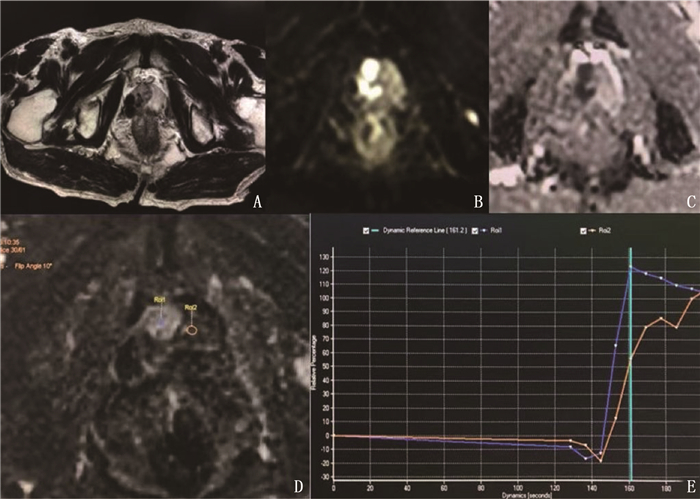
恶性组患者的容量转移常数(Ktrans)、速率常数(Kep)、血管外细胞外间隙容积比(Ve)大于良性组,差异有统计学意义(P < 0.05), 见表 1。前列腺恶性病变患者MRI图像见图 1。
表 1 前列腺良恶性病变患者的DCE-MRI指标比较(x±s)组别 n Ktrans/min Kep/min Ve 良性组 65 0.68±0.08 0.61±0.05 0.64±0.04 恶性组 37 0.73±0.06* 0.67±0.06* 0.68±0.05* 与良性组比较, * P < 0.05。 2.2 前列腺良恶性病变患者的SI-T曲线比较
前列腺良恶性病变患者的SI-T曲线类型比较,差异有统计学意义(P < 0.05)。恶性组患者的SI-T曲线速升缓降型占比高于良性组,持续缓升型、缓升平台型占比低于良性组,差异有统计学意义(P < 0.05), 见表 2。
表 2 前列腺良恶性病变患者的SI-T曲线比较[n(%)]组别 n 持续缓升型 缓升平台型 速升平台型 速升缓降型 良性组 65 19(29.23) 23(35.38) 18(27.69) 5(7.69) 恶性组 37 2(5.41)* 5(13.51)* 12(32.43) 18(48.65)* 与良性组比较, * P < 0.05。 2.3 前列腺良恶性病变患者的SI-T曲线参数比较
恶性组患者的达峰时间短于良性组,强化程度、强化率高于良性组,差异有统计学意义(P < 0.05), 见表 3。
表 3 前列腺良恶性病变患者的SI-T曲线参数比较(x±s)组别 n 达峰时间/s 强化程度/% 强化率/% 良性组 65 126.27±20.49 153.05±10.31 18.15±4.92 恶性组 37 101.15±18.57* 160.89±9.04* 24.71±5.08* 与良性组比较, * P < 0.05。 2.4 DCE-MRI指标及SI-T曲线参数对前列腺良恶性病变的诊断价值
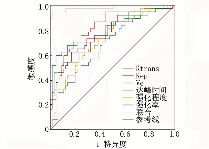
联合检测诊断前列腺良恶性病变的曲线下面积(AUC)大于DCE-MRI指标及SI-T曲线参数单独检测,差异有统计学意义(P < 0.05), 见表 4、图 2。
表 4 DCE-MRI指标及SI-T曲线参数对前列腺良恶性病变的诊断价值指标 截点值 AUC SE 95%CI 敏感度/% 特异度/% DCE-MRI指标 Ktrans 0.72 min 0.693* 0.053 0.590~0.796 58.46 70.27 Kep 0.64 min 0.777* 0.049 0.681~0.873 83.08 65.16 Ve 0.68 0.757* 0.051 0.657~0.857 86.15 54.05 SI-T曲线 达峰时间 119.59 s 0.828* 0.043 0.743~0.913 64.62 86.49 强化程度 152.58% 0.731* 0.051 0.631~0.830 49.23 86.49 强化率 23.31% 0.806* 0.049 0.711~0.902 95.38 59.46 联合检测 0.982 0.014 0.954~1.000 98.46 94.59 Ktrans: 容量转移常数; Kep: 速率常数; Ve: 血管外细胞外间隙容积比。与联合检测比较, * P < 0.05。 3. 讨论
前列腺癌指前列腺上皮性恶性肿瘤,是中老年男性常见恶性肿瘤疾病,随着社会老龄化程度加剧,前列腺癌发生率呈升高趋势[5-6]。既往研究[7-8]发现,常规超声、磁共振成像(MRI)等难以准确鉴别前列腺癌及良性病变,故需探究一种诊断准确率高的检查方法。DCE-MRI是一种常见的检查方法,主要基于病变区血供多少、血管通透性及细胞外间隙大小等参数进行检查,可反映新生血管特性[9-11]。本研究结果显示,恶性病变患者的Ktrans、Kep、Ve值大于良性病变患者,即前列腺恶性病变患者新生血管形成较为旺盛,与相关研究结果相符。
DCE-MRI在组织和肿瘤血管生理特性的评估中表现出无创性成像的优势,在静脉团注对比剂后,可显示组织、血管中浓度的变化,进而可观察到肿瘤新生血管通透性的改变、肿瘤血管生成[12-14]。SI-T曲线是随时间而变化的DCE-MRI信号,可反映ROI组织造影剂廓清特性。相关研究[15-17]指出,达峰时间、强化率等SI-T曲线指标可较好地反映前列腺组织微循环特点。达峰时间主要反映对比剂流入组织的速度。前列腺癌血管密度增加,新生毛细血管基底膜不连续、管径粗细不均及管壁缺乏肌层和基底膜,导致血管通透性增加,对比剂廓清较快,故早期强化明显[18-20]。本研究结果显示,恶性病变患者的达峰时间短于良性病变患者,强化程度、强化率高于良性病变患者,表明恶性病变存在DCE-MRI早期强化现象,这主要是因为前列腺癌组织血管较为丰富,新生血管不成熟,肿瘤血管内皮不完整,血管壁通透性增加,血管阻力较小,MRI动态增强扫描表现为达峰时间明显缩短,强化率增加,呈现为明显早期强化等特点[21-23]。此外,本研究结果显示,联合检测诊断前列腺良恶性病变的AUC大于DCE-MRI指标及SI-T曲线参数单独检测,表明联合检查对前列腺良恶性病具有较好的诊断价值,提示或可用DCE-MRI对前列腺病变进行早期诊断。
临床研究[24]显示,恶性肿瘤的生长、浸润及转移均依赖于新生血管生成,且新生肿瘤微血管的形态及功能均不同于正常血管。肿瘤微血管通透性增加,血管内皮细胞间隙及血管流动性也与正常血管有较大差异。SI-T曲线是一种能对肿瘤微循环生理功能状态进行“成像”的影像学方法,能得到肿瘤强化过程中更精确的信息,可量化反映病变组织血供情况。研究[25]表明,SI-T曲线测量参数与肿瘤血管生成功能高度相关。临床资料[26]显示,前列腺良恶性病变组织新生血管较正常前列腺组织丰富,在进行DCE-MRI扫描时,表现为早期明显强化现象,而前列腺增生组织血管密度较大,血管腔内对比剂难以进行组织间隙,故在SI-T曲线中表现为早期强化后持续性增高,或出现平台期。本研究发现,前列腺良性病变中持续缓升型、缓升平台型较多,且前列腺癌患者中SI-T曲线速升平台型、速升缓降型检出率较高,这主要与癌组织中的血管分布不均、粗细不均及新生血管壁不成熟、血管壁基底膜不连续、血管通透性增高有关。
综上所述, DCE-MRI及SI-T曲线联合检查对前列腺良恶性病变具有较好的诊断价值。
-
表 1 前列腺良恶性病变患者的DCE-MRI指标比较(x±s)
组别 n Ktrans/min Kep/min Ve 良性组 65 0.68±0.08 0.61±0.05 0.64±0.04 恶性组 37 0.73±0.06* 0.67±0.06* 0.68±0.05* 与良性组比较, * P < 0.05。 表 2 前列腺良恶性病变患者的SI-T曲线比较[n(%)]
组别 n 持续缓升型 缓升平台型 速升平台型 速升缓降型 良性组 65 19(29.23) 23(35.38) 18(27.69) 5(7.69) 恶性组 37 2(5.41)* 5(13.51)* 12(32.43) 18(48.65)* 与良性组比较, * P < 0.05。 表 3 前列腺良恶性病变患者的SI-T曲线参数比较(x±s)
组别 n 达峰时间/s 强化程度/% 强化率/% 良性组 65 126.27±20.49 153.05±10.31 18.15±4.92 恶性组 37 101.15±18.57* 160.89±9.04* 24.71±5.08* 与良性组比较, * P < 0.05。 表 4 DCE-MRI指标及SI-T曲线参数对前列腺良恶性病变的诊断价值
指标 截点值 AUC SE 95%CI 敏感度/% 特异度/% DCE-MRI指标 Ktrans 0.72 min 0.693* 0.053 0.590~0.796 58.46 70.27 Kep 0.64 min 0.777* 0.049 0.681~0.873 83.08 65.16 Ve 0.68 0.757* 0.051 0.657~0.857 86.15 54.05 SI-T曲线 达峰时间 119.59 s 0.828* 0.043 0.743~0.913 64.62 86.49 强化程度 152.58% 0.731* 0.051 0.631~0.830 49.23 86.49 强化率 23.31% 0.806* 0.049 0.711~0.902 95.38 59.46 联合检测 0.982 0.014 0.954~1.000 98.46 94.59 Ktrans: 容量转移常数; Kep: 速率常数; Ve: 血管外细胞外间隙容积比。与联合检测比较, * P < 0.05。 -
[1] CHANG Y X, DENG Q, GUAN Z F, et al. miR-1273g-3p promotes malignant progression and has prognostic implications in prostate cancer[J]. Mol Biotechnol, 2022, 64(1): 17-24. doi: 10.1007/s12033-021-00384-x
[2] 余英芳, 胡海霞, 王小义, 等. 动态对比增强MRI在肺良恶性结节(≤2 cm)的诊断价值[J]. 中国CT和MRI杂志, 2022, 20(11): 64-66. https://www.cnki.com.cn/Article/CJFDTOTAL-CTMR202211024.htm [3] 刘晓东, 方习奇, 唐桑, 等. 动态对比增强MRI联合表观扩散系数值对前列腺中央区癌的诊断价值[J]. 实用放射学杂志, 2020, 36(4): 599-602. [4] 中国抗癌协会泌尿男生殖系肿瘤专业委员会微创学组. 中国前列腺癌外科治疗专家共识[J]. 浙江医学, 2018, 40(3): 217-220. https://www.cnki.com.cn/Article/CJFDTOTAL-ZGAZ202212013.htm [5] ALLARAKHA A, GAO Y, JIANG H, et al. Predictive ability of DWI/ADC and DCE-MRI kinetic parameters in differentiating benign from malignant breast lesions and in building a prediction model[J]. Discov Med, 2019, 27(148): 139-152.
[6] NAKANISHI K, TANAKA J, NAKAYA Y, et al. Whole-body MRI: detecting bone metastases from prostate cancer[J]. Jpn J Radiol, 2022, 40(3): 229-244. doi: 10.1007/s11604-021-01205-6
[7] 孙文杰, 王欣, 刘玲, 等. 3. 0T磁共振多参数成像及动态增强扫描对前列腺癌的诊断价值[J]. 中国CT和MRI杂志, 2022, 20(1): 138-141. https://www.cnki.com.cn/Article/CJFDTOTAL-CTMR202201043.htm [8] 任义财, 周利平. DCE-MRI、3D1H-MRS联合对前列腺良恶性病变的诊断价值[J]. 中国CT和MRI杂志, 2020, 18(4): 107-109, 113. https://www.cnki.com.cn/Article/CJFDTOTAL-CTMR202004032.htm [9] 杜元元, 李玉智, 马蔚, 等. DWI、1H-MRS及DCE-MR对前列腺良恶性病变的诊断价值[J]. 海南医学, 2022, 33(3): 352-356. https://www.cnki.com.cn/Article/CJFDTOTAL-HAIN202203020.htm [10] 贾海云, 王佳强, 周晓莹, 等. 剪切波弹性成像联合多参数MRI对临床显著性前列腺癌的诊断价值[J]. 临床超声医学杂志, 2022, 24(8): 603-607. https://www.cnki.com.cn/Article/CJFDTOTAL-LCCY202208008.htm [11] 李鹏, 黄英, 李艳, 等. DWI和DCE-MRI鉴别诊断良恶性前列腺外周带T2WI局灶性低信号病变[J]. 中国介入影像与治疗学, 2021, 18(3): 156-160. https://www.cnki.com.cn/Article/CJFDTOTAL-JRYX202103011.htm [12] 刘郭坤, 李健斐, 刘艳超, 等. IVIM-DWI与DCE-MRI在前列腺疾病诊断中的联合应用观察[J]. 山东医药, 2020, 60(3): 75-77. https://www.cnki.com.cn/Article/CJFDTOTAL-SDYY202003023.htm [13] STABILE A, BARLETTA F, MOTTERLE G, et al. Optimizing prostate-targeted biopsy schemes in men with multiple mpMRI visible lesions: should we target all suspicious areas Results of a two institution series[J]. Prostate Cancer Prostatic Dis, 2021, 24(4): 1137-1142. doi: 10.1038/s41391-021-00371-y
[14] MONTORSI F, STABILE A, GANDAGLIA G, et al. Re: Marra etal. 'Transperineal freehand multiparametric MRI fusion targeted biopsies under local anaesthesia for prostate cancer diagnosis: a multicentre prospective study of 1014 cases'[J]. BJU Int, 2021, 128(4): 523.
[15] 刘凯, 罗红兰, 柯楠, 等. DCE-MRI和IVIM-DWI在前列腺癌病理分级和临床分期中的诊断价值分析[J]. 中国性科学, 2022, 31(7): 31-35. https://www.cnki.com.cn/Article/CJFDTOTAL-XKXZ202207009.htm [16] 陈爽, 陈子健, 钟建锋, 等. 动态对比增强磁共振联合PSA诊断前列腺癌价值探讨[J]. 中国CT和MRI杂志, 2022, 20(1): 142-145. https://www.cnki.com.cn/Article/CJFDTOTAL-CTMR202201044.htm [17] GASSENMAIER S, AFAT S, NICKEL D, et al. Deep learning-accelerated T2-weighted imaging of the prostate: reduction of acquisition time and improvement of image quality[J]. Eur J Radiol, 2021, 137: 109600.
[18] 邰兆琴, 徐小虎, 许亚春, 等. 磁共振动态增强联合DWI与超声引导穿刺对照在前列腺病变诊断中的应用[J]. 中国CT和MRI杂志, 2021, 19(8): 113-116. https://www.cnki.com.cn/Article/CJFDTOTAL-CTMR202108037.htm [19] WEI C G, CHEN T, ZHANG Y Y, et al. Erratum to"Biparameteric prostate MRI and clinical indicators predict clinically significant prostate cancer in men with" gray zone "PSA levels"[J]. Eur J Radiol, 2020, 129: 109129.
[20] 李珍, 邹磊, 弥长虹, 等. 前列腺癌磁共振动态增强定量研究[J]. 中国CT和MRI杂志, 2021, 19(7): 126-127, 137. https://www.cnki.com.cn/Article/CJFDTOTAL-CTMR202107036.htm [21] PESAPANE F, STANDAERT C, VISSCHERE P D, et al. T-staging of prostate cancer: identification of useful signs to standardize detection of posterolateral extraprostatic extension on prostate MRI[J]. Clin Imaging, 2020, 59(1): 1-7.
[22] 陈莉, 黄道煌, 骆祥伟, 等. 3. 0T磁共振动态增强定量参数联合DWI诊断前列腺癌临床价值[J]. 医学影像学杂志, 2022, 32(6): 1073-1075. https://www.cnki.com.cn/Article/CJFDTOTAL-XYXZ202206043.htm [23] STONE L. Piloting hyperpolarized 13C-pyruvate MRI for imaging advanced prostate cancer[J]. Nat Rev Urol, 2020, 17(1): 7.
[24] PARRA N A, LU H, LI Q, et al. Erratum: predicting clinically significant prostate cancer using DCE-MRI habitat descriptors[J]. Oncotarget, 2019, 10(21): 2113.
[25] 李盈盈, 张明博, 吴凡, 等. 经直肠多模态超声与多模态MRI检查对前列腺癌诊断价值的对照研究[J]. 中华泌尿外科杂志, 2020, 41(1): 19-25. https://cpfd.cnki.com.cn/Article/CPFDTOTAL-CSYX201810001237.htm [26] CHRISTOPHE C, MONTAGNE S, BOURRELIER S, et al. Prostate cancer local staging using biparametric MRI: assessment and comparison with multiparametric MRI[J]. Eur J Radiol, 2020, 132: 109350.
-
期刊类型引用(4)
1. 承伟涓. 动态对比增强磁共振鉴别诊断前列腺癌和良性前列腺增生的效果观察. 影像研究与医学应用. 2024(04): 57-59 .  百度学术
百度学术
2. 彭强. 特异性标志物免疫组化在前列腺良、恶性病变诊断中的价值研究. 慢性病学杂志. 2024(05): 707-710 .  百度学术
百度学术
3. 曾勇,蒋中灿. MRI动态增强扫描在乳腺肿瘤性疾病的诊断价值. 现代医用影像学. 2024(06): 1065-1067+1071 .  百度学术
百度学术
4. 丁云云,张继良,武东杰. 动态增强磁共振成像联合血清游离前列腺特异性抗原检测对前列腺癌的诊断价值. 航空航天医学杂志. 2024(09): 1041-1044 .  百度学术
百度学术
其他类型引用(0)


 下载:
下载:


 苏公网安备 32100302010246号
