
Mining of core genes and analysis of related pathways in Alzheimer's disease based on bioinformatic analysis
-
摘要:目的
基于生物信息学分析筛选阿尔茨海默病(AD)的核心基因,并对可能的致病信号通路进行分析。
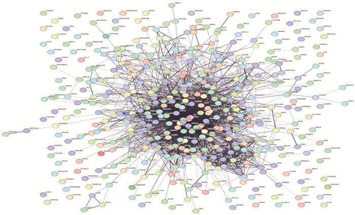
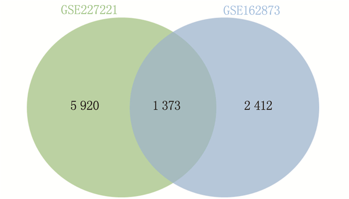
方法从基因表达综合(GEO)数据库中下载AD相关基因芯片数据,筛选出GSE227221和GSE162873数据集。应用GEO2R在线分析软件筛选AD组织样本和正常脑组织样本间的差异表达基因(DEGs)。应用R软件DOSE包对DEGs进行疾病本体论(DO)基因富集分析,使用在线数据分析工具DAVID对DEGs进行基因本体论(GO)和京都基因与基因组百科全书(KEGG)富集分析。通过STRING数据库分析基因的蛋白质交互作用,应用Cytoscape软件构建蛋白质-蛋白质相互作用(PPI)网络,并通过MCODE插件对PPI网络进行枢纽基因的分析与筛选。根据MCODE评分,对枢纽基因进一步分析并筛选出核心基因。
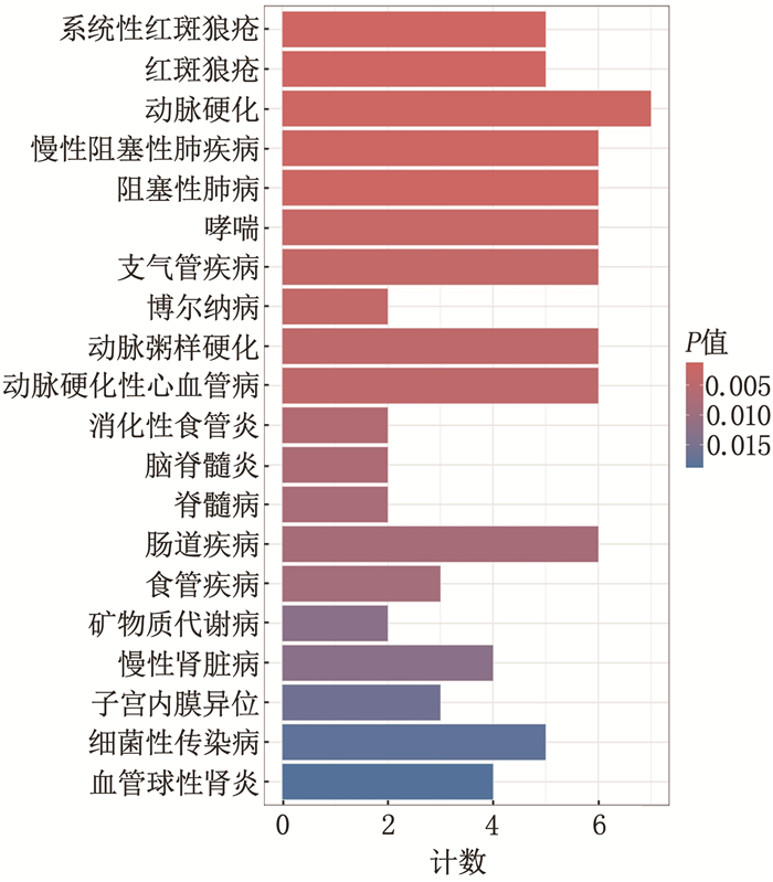
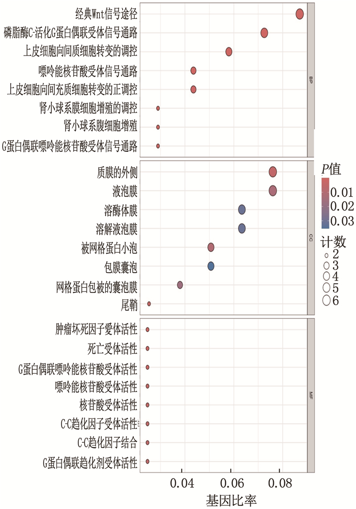
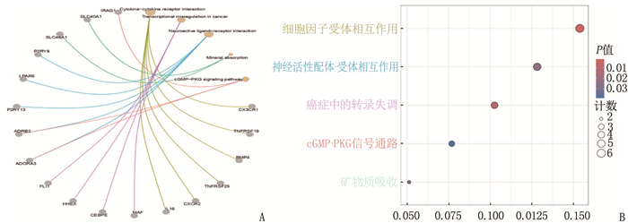
结果对2个数据集进行比对后,共筛选出参与AD发生和进展的1 373个相同变化趋势的DEGs, 并筛选验证出核心基因为GIMAP基因(包括GIMAP1、GIMAP4、GIMAP5、GIMAP6、GIMAP7及GIMAP1-GIMAP5)。DO基因富集分析结果显示, DEGs与系统性红斑狼疮、红斑狼疮和动脉硬化这3种疾病最为相关; GO功能富集分析结果显示, DEGs主要富集于3条信号通路,即经典Wnt信号通路、磷脂酶C-活化G蛋白质-耦合受体通路和等离子体外侧膜通路; KEGG信号通路富集分析结果显示, DEGs主要富集于细胞因子-细胞因子受体相互作用、神经元活性配体-受体相互作用、癌症相关的转录失调等通路。
结论AD的核心致病基因可能为GIMAP基因,与AD发生相关的通路复杂,可能与免疫功能紊乱有关。
Abstract:ObjectiveTo identify core genes of Alzheimer's disease (AD) through bioinformatic analysis and explore potential pathogenic signaling pathways.
MethodsGene expression profiling data related to AD were downloaded from the Gene Expression Omnibus (GEO) database, with datasets GSE227221 and GSE162873 selected for analysis. GEO2R online analysis software was applied to screen differentially expressed genes (DEGs) between AD tissue samples and normal brain tissue samples. The Disease Ontology (DO) enrichment analysis of DEGs was performed using the DOSE package in R software, while Gene Ontology (GO) and Kyoto Encyclopedia of Genes and Genomes (KEGG) enrichment analyses were conducted through the online data analysis tool DAVID. Protein-protein interactions (PPI) among genes were analyzed using the STRING database, and a PPI network was constructed with Cytoscape software. Hub genes within the PPI network were identified and screened using the MCODE plugin. Based on MCODE scores, further analysis was conducted to select core genes.
ResultsA total of 1, 373 DEGs with consistent expression trends involved in the onset and progression of AD were identified from the two datasets, with the core genes of GIMAP genes (including GIMAP1, GIMAP4, GIMAP5, GIMAP6, GIMAP7, and GIMAP1-GIMAP5). DO enrichment analysis revealed the strongest associations between DEGs and three diseases: systemic lupus erythematosus, lupus erythematosus, and atherosclerosis. GO functional enrichment analysis indicated that DEGs were primarily enriched in three signaling pathways: the classical Wnt signaling pathway, phospholipase C-activated G protein-coupled receptor pathway, and plasma membrane lateral domain pathway. KEGG pathway enrichment analysis showed that DEGs were primarily enriched in pathways such as cytokine-cytokine receptor interaction, neuroactive ligand-receptor interaction, and cancer-related transcriptional dysregulation.
ConclusionThe core pathogenic gene of AD may be the GIMAP gene, and the associated pathways are complex, potentially implicating immune dysfunction.
-
-
[1] RAJESH Y, KANNEGANTI T D. Innate immune cell death in neuroinflammation and Alzheimer's disease[J]. Cells, 2022, 11(12): 1885. doi: 10.3390/cells11121885
[2] SUN N, VICTOR M B, PARK Y P, et al. Human microglial state dynamics in Alzheimer's disease progression[J]. Cell, 2023, 186(20): 4386-4403. doi: 10.1016/j.cell.2023.08.037
[3] HANNA R, FLAMIER A, BARABINO A, et al. G-quadruplexes originating from evolutionary conserved L1 elements interfere with neuronal gene expression in Alzheimer's disease[J]. Nat Commun, 2021, 12(1): 1828. doi: 10.1038/s41467-021-22129-9
[4] CROUS-BOU M, MINGUILL N C, GRAMUNT N, et al. Alzheimer's disease prevention: from risk factors to early intervention[J]. Alzheimers Res Ther, 2017, 9(1): 71. doi: 10.1186/s13195-017-0297-z
[5] THIES W, BLEILER L. 2021 Alzheimer's disease facts and figures[J]. Alzheimers Dement, 2021, 17(3): 327-406. doi: 10.1002/alz.12328
[6] LONG J M, HOLTZMAN D M. Alzheimer disease: an update on pathobiology and treatment strategies[J]. Cell, 2019, 179(2): 312-339. doi: 10.1016/j.cell.2019.09.001
[7] PANZA F, LOZUPONE M, LOGROSCINO G, et al. A critical appraisal of amyloid-β-targeting therapies for Alzheimer disease[J]. Nat Rev Neurol, 2019, 15(2): 73-88. doi: 10.1038/s41582-018-0116-6
[8] KRÜCKEN J, SCHROETEL R M, MÜLLER I U, et al. Comparative analysis of the human gimap gene cluster encoding a novel GTPase family[J]. Gene, 2004, 341: 291-304. doi: 10.1016/j.gene.2004.07.005
[9] NITTA T, TAKAHAMA Y. The lymphocyte guard-IANs: regulation of lymphocyte survival by IAN/GIMAP family proteins[J]. Trends Immunol, 2007, 28(2): 58-65. doi: 10.1016/j.it.2006.12.002
[10] NITTA T, NASREEN M, SEIKE T, et al. IAN family critically regulates survival and development of T lymphocytes[J]. PLoS Biol, 2006, 4(4): e103. doi: 10.1371/journal.pbio.0040103
[11] GROSCH H. Subcortical dementia with pseudofocal motor disorders: mental deficiency in a 9-year old girl after whooping cough vaccination[J]. Dtsch Z Nervenheilkd, 1953, 170(3): 237-253. doi: 10.1007/BF00218543
[12] SOLITARE G B. Neuronophagia[J]. Lancet, 1979, 1(8112): 387.
[13] YE Y, GAO M Z, SHI W T, et al. The immunomodulatory effects of mesenchymal stem cell-derived extracellular vesicles in Alzheimer's disease[J]. Front Immunol, 2023, 14: 1325530.
[14] KRÜCKEN J, SCHROETEL R M, MÜLLER I U, et al. Comparative analysis of the human gimap gene cluster encoding a novel GTPase family[J]. Gene, 2004, 341: 291-304. doi: 10.1016/j.gene.2004.07.005
[15] ZENZ T, ROESSNER A, THOMAS A, et al. hIan5: the human ortholog to the rat Ian4/Iddm1/lyp is a new member of the Ian family that is overexpressed in B-cell lymphoid malignancies[J]. Genes Immun, 2004, 5(2): 109-116. doi: 10.1038/sj.gene.6364044
[16] BARNES M J, AKSOYLAR H, KREBS P, et al. Loss of T cell and B cell quiescence precedes the onset of microbial flora-dependent wasting disease and intestinal inflammation in Gimap5-deficient mice[J]. J Immunol, 2010, 184(7): 3743-3754. doi: 10.4049/jimmunol.0903164
[17] SCHULTEIS R D, CHU H Y, DAI X Z, et al. Impaired survival of peripheral T cells, disrupted NK/NKT cell development, and liver failure in mice lacking Gimap5[J]. Blood, 2008, 112(13): 4905-4914. doi: 10.1182/blood-2008-03-146555
[18] DATTA P, WEBB L M, AVDO I, et al. Survival of mature T cells in the periphery is intrinsically dependent on GIMAP1 in mice[J]. Eur J Immunol, 2017, 47(1): 84-93. doi: 10.1002/eji.201646599
[19] WEBB L M, DATTA P, BELL S E, et al. GIMAP1 is essential for the survival of naive and activated B cells in vivo[J]. J Immunol, 2016, 196(1): 207-216. doi: 10.4049/jimmunol.1501582
[20] SCHNELL S, DÉMOLLIōRE C, VAN DEN BERK P, et al. Gimap4 accelerates T-cell death[J]. Blood, 2006, 108(2): 591-599. doi: 10.1182/blood-2005-11-4616
[21] HO C H, TSAI S F. Functional and biochemical characterization of a T cell-associated anti-apoptotic protein, GIMAP6[J]. J Biol Chem, 2017, 292(22): 9305-9319. doi: 10.1074/jbc.M116.768689
[22] DAI P, RUAN P L, MAO Y, et al. Gimap5 promoted RSV degradation through interaction with M6PR[J]. J Med Virol, 2023, 95(1): e28390. doi: 10.1002/jmv.28390
[23] XIE T H, LIU X, LI P. CD138 promotes the accumulation and activation of autoreactive T cells in autoimmune MRL/lpr mice[J]. Exp Ther Med, 2023, 26(6): 568. doi: 10.3892/etm.2023.12267
[24] ZHANG M, LI J X, HUA C C, et al. Exploring an immune cells-related molecule in STEMI by bioinformatics analysis[J]. BMC Med Genomics, 2023, 16(1): 151. doi: 10.1186/s12920-023-01579-8
[25] MARQUES E R M D C, HSIEH R, LOURENÇO S V, et al. Oral lupus erythematosus: Immunohistochemical evaluation of CD1a, CD21, CD123, and langerin expression in dendritic cells[J]. J Cutan Pathol, 2024, 51(5): 368-378. doi: 10.1111/cup.14568
 下载:
下载:

计量
- 文章访问数: 168
- HTML全文浏览量: 22
- PDF下载量: 30
 苏公网安备 32100302010246号