| Citation: |
XIONG Chaoyue, ZHOU Min, HE Xiaofang, HUA Xiaofan. Value of prediction model established based on ultrasound features combined with clinical data in evaluating axillary lymph node metastasis in patients with early breast cancer[J]. Journal of Clinical Medicine in Practice, 2022, 26(12): 14-18, 22. DOI: 10.7619/jcmp.20220195

|
To explore value of prediction model established based on ultrasound features combined with clinical data in evaluating axillary lymph node (ALN) metastasis in patients with early breast cancer.
The ultrasonic characteristics and clinical data of 203 women with unilateral early breast cancer were retrospectively analyzed. The patients were divided into metastatic group and non-metastatic group, and were determined whether they had ALN metastasis or not according to the pathological results. Single factor screening was performed for each index of the two groups. Logistic multivariate regression analysis was performed again and a prediction model was established. Receiver operating characteristic (ROC) curve was used to detect its discrimination, and goodness-of-fit test was used to evaluate the degree of calibration. Another 78 unilateral early breast cancer patients in our hospital were selected for clinical validation of the model.
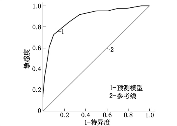
The pathological results of 203 breast cancer patients showed that ALN metastasis occurred in 71 cases(metastasis group), accounting for 34.98%. Tumor diameter ≥3 cm, blurred tumor margin, longer ALN short diameter, higher ALN short diameter to long diameter ratio, higher value of ALN cortical thickness, lower degree of differentiation, and higher level of serum microRNA-21(miRNA-21) expression were all risk factors for ALN metastasis in breast cancer patients (P<0.05). According to the risk factors, the prediction model expression equation was as follows. Logit(P)=1.912×tumor diameter ≥3 cm(yes=1, no=0)+2.040×tumor margin blur(yes=1, no=0)+1.582×ALN short diameter(measured value)+3.374×ALN short/long diameter(measured value)+2.264×ALN cortical thickness(measured value)+2.497×differentiation degree(yes=1, no=0)+2.921×miRNA-21 expression amount(measured value)-33.615. The area of the ROC curve of this model was 0.886 (95%CI, 0.838 to 0.933), the sensitivity and specificity corresponding to the maximum Youden index (0.736) were 88.50% and 83.60% respectively. Goodness of fit test showed that the model did not overfit (χ2=2.067, P=0.394). Clinical validation results showed that the sensitivity of the model was 87.10%, the specificity was 82.98%, and the accuracy was 84.62%.
It is valuable in predicting the risk of ALN metastasis by constructing a predictive model based on degree of tumor differentiation, serum miRNA-21 expression, tumor diameter, tumor margin, and ALN short-diameter, short-diameter/long-diameter ratio, and cortical thickness in early breast cancer patients.
| [1] |
BUNDRED N J, BARNES N L P, RUTGERS E, et al. Is axillary lymph node clearance required in node-positive breast cancer[J]. Nat Rev Clin Oncol, 2015, 12(1): 55-61. doi: 10.1038/nrclinonc.2014.188
|
| [2] |
JOO J H, KIM S S, SON B H, et al. Evaluation of the prognostic stage in the 8th edition of the American joint committee on cancer in patients with breast cancer and internal mammary lymph node metastasis[J]. Anticancer Res, 2018, 38(9): 5357-5361. doi: 10.21873/anticanres.12864
|
| [3] |
中国抗癌协会乳腺癌专业委员会. 中国抗癌协会乳腺癌诊治指南与规范(2011版)[J]. 中国癌症杂志, 2011, 21(5): 367-417. doi: 10.3969/j.issn.1007-3639.2011.05.010
|
| [4] |
牛昀. 2012年版《WHO乳腺肿瘤分类》新变化与临床治疗的关系[J]. 中华乳腺病杂志: 电子版, 2014, 8(3): 156-160. https://www.cnki.com.cn/Article/CJFDTOTAL-ZHRD201403002.htm
|
| [5] |
周子君, 古林, 张乃千, 等. T1a~1b期老年乳腺癌腋窝淋巴结转移的相关影响因素[J]. 中国老年学杂志, 2021, 41(19): 4209-4211. doi: 10.3969/j.issn.1005-9202.2021.19.016
|
| [6] |
王宋, 权毅. Luminal A型乳腺癌发生腋窝淋巴结转移的风险因素分析[J]. 中国普外基础与临床杂志, 2021, 28(6): 789-793. https://www.cnki.com.cn/Article/CJFDTOTAL-ZPWL202106019.htm
|
| [7] |
BLVTHNER E, BEDNARSCH J, MALINOWSKI M, et al. Dynamic liver function is an independent predictor of recurrence-free survival after curative liver resection for HCC-A retrospective cohort study[J]. Int J Surg, 2019, 71: 56-65. doi: 10.1016/j.ijsu.2019.08.033
|
| [8] |
刘信礼, 王雯, 牛学才, 等. 早期浸润性乳腺癌患者腋窝淋巴结转移的影响因素分析[J]. 山东医药, 2021, 61(28): 62-65. https://www.cnki.com.cn/Article/CJFDTOTAL-SDYY202128015.htm
|
| [9] |
TSENG H S, CHEN L S, KUO S J, et al. Tumor characteristics of breast cancer in predicting axillary lymph node metastasis[J]. Med Sci Monit, 2014, 20: 1155-1161. doi: 10.12659/MSM.890491
|
| [10] |
ANDERSSON Y, BERGKVIST L, FRISELL J, et al. Long-term breast cancer survival in relation to the metastatic tumor burden in axillary lymph nodes[J]. Breast Cancer Res Treat, 2018, 171(2): 359-369. doi: 10.1007/s10549-018-4820-0
|
| [11] |
SUSINI T, NORI J, OLIVIERI S, et al. Predicting the status of axillary lymph nodes in breast cancer: a multiparameter approach including axillary ultrasound scanning[J]. Breast, 2009, 18(2): 103-108. doi: 10.1016/j.breast.2009.02.001
|
| [12] |
YILMAZ M H, ESEN G, AYARCAN Y, et al. The role of US and MR imaging in detecting local chest wall tumor recurrence after mastectomy[J]. Diagn Interv Radiol, 2007, 13(1): 13-18.
|
| [13] |
WU J L, TSENG H S, YANG L H, et al. Prediction of axillary lymph node metastases in breast cancer patients based on pathologic information of the primary tumor[J]. Med Sci Monit, 2014, 20: 577-581. doi: 10.12659/MSM.890345
|
| [14] |
许睿, 钱军, 张明亮, 等. 长链非编码RNA MEG3对乳腺癌细胞侵袭及迁移的影响[J]. 赣南医学院学报, 2021, 41(3): 256-260. https://www.cnki.com.cn/Article/CJFDTOTAL-GNYX202103008.htm
|
| [15] |
ZHANG C F, LIU K, LI T, et al. miR-21: a gene of dual regulation in breast cancer[J]. Int J Oncol, 2016, 48(1): 161-172. doi: 10.3892/ijo.2015.3232
|
| [16] |
张瑞瑞, 赵桓玉, 周武碧, 等. 乳腺癌组织中Bmi-1和CARD9表达及其与淋巴结转移和预后的关联[J]. 西部医学, 2021, 33(11): 1642-1646, 1659. https://www.cnki.com.cn/Article/CJFDTOTAL-XIBU202111019.htm
|
| [17] |
LAMPIS A, CAROTENUTO P, VLACHOGIANNIS G, et al. MIR21 drives resistance to heat shock protein 90 inhibition in cholangiocarcinoma[J]. Gastroenterology, 2018, 154(4): 1066-1079. e5. doi: 10.1053/j.gastro.2017.10.043
|
| [18] |
唐诗聪, 陈东, 郭瑢, 等. miRNA-21调控HER-2阳性乳腺癌的血管生成[J]. 昆明医科大学学报, 2020, 41(8): 39-45. https://www.cnki.com.cn/Article/CJFDTOTAL-KMYX202008007.htm
|
| 1. |
刘文通. 甲状腺功能5项对鉴别诊断甲状腺功能亢进和甲状腺功能减退中的价值研究. 黑龙江中医药. 2024(01): 359-361 .

| |
| 2. |
蒋瑞妹,王卓群,牛敏,申金付,秦瑶,李娟. 骨吸收标志物β-CTX与Graves病患者并发高钙血症的相关性. 中国医师杂志. 2023(04): 528-531, 536 .
| |
| 3. |
祖力皮亚·努尔买买提,热依汗尼沙·亚克亚,阿不都力木·斯迪克. 普萘洛尔辅助丙硫氧嘧啶治疗老年甲状腺亢进的疗效及对糖脂代谢和ACTH水平的影响. 中外医疗. 2023(23): 92-95 .
| |
| 4. |
乔辉,丁红娜,孙君. 甲状腺功能亢进患者骨密度、骨代谢、脂肪细胞因子的相关性. 河南医学研究. 2023(22): 4091-4095 .
| |
| 5. |
郭小妮,刘师伟,段瑞雪,赵宇翔,吴亚茹. 脂肪因子vaspin与骨质疏松症相关性的研究进展. 生命的化学. 2022(10): 1905-1912 .
|
© 2020 《实用临床医药杂志》编辑部
Address: 江苏省扬州市江阳中路136号,扬州大学江阳路北校区14号楼201室China Pos: 225009Tel: 0514-87978917、87978989、87978807
Supported by:
Beijing Renhe Information Technology Co., Ltd.

 苏公网安备 32100302010246号
 DownLoad: