| Citation: |
LIU Bo, CAO Guanghua, ZHANG Wenxi, YANG Dong, JIANG Hui, QIAO Zhijun. Effect observation of 3D printing adjuvant therapy in treatment of patients with posterior Pilon fracture after treatment[J]. Journal of Clinical Medicine in Practice, 2022, 26(17): 10-14. DOI: 10.7619/jcmp.20221193

|
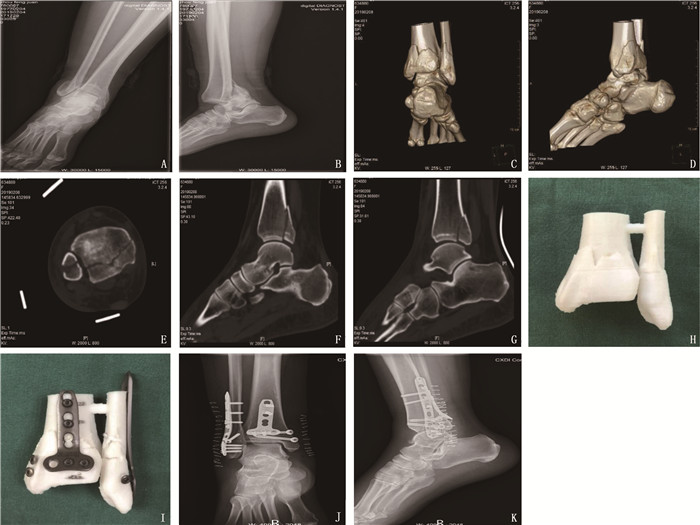
To observe the clinical effect of 3D printing adjuvant therapy in treatment of patients with posterior Pilon fracture after treatment.
The clinical materials of 92 patients with posterior Pilon fracture from January 2012 to December 2020 in the Department of Orthopedics of Liyang People′s Hospital were retrospectively analyzed. Among them, 47 patients with preoperative 3D printing for simulating operation were selected as 3D printing group, and 45 patients without 3D printing for simulating operation were selected as control group. The operation related complications were recorded in both groups, and the operation time, intraoperative X-ray fluoroscopy times, condition of surgical reduction, clinical healing time of fractures, the score of ankle scoring system of the American College of Foot and Ankle Surgeons (AOFAS) and the score of Visual Analogue Scale (VAS) of ankle pain at the last follow-up were compared between the two groups.
All the patients were followed up for 12 to 20 months. All the patients achieved primary healing after operation, and no patient had complications such as incision infection, skin necrosis, deep venous thrombosis of lower limbs, and checkrein deformity of toes. The operation time and intraoperative fluoroscopy times in the 3D printing group were (81.4±9.4) min and (13.0±2.3) times respectively, which were significantly shorter and less than (94.9±11.6) min and (18.4±3.2) times in the control group (P < 0.05). In the control group, there were 3 cases with poor reduction of posterior malleolus fracture, but there were no patients with poor reduction of posterior malleolus fracture in the 3D printing group, and there was no significant difference between two groups (P>0.05). The fracture healing time, postoperative AOFAS score and VAS score in the 3D printing group were (12.9±1.0) weeks, (92.8±4.2) points and (1.1±0.8) points respectively, while were (13.2±1.3) weeks, (90.8±4.5) points and (1.3±0.8) points respectively in the control group, and there were no significant differences between two groups (P>0.05).
Application of 3D printing adjuvant therapy can optimize the operation plan of posterior Pilon fracture, shorten the operation time, reduce the number of intraoperative fluoroscopy, and achieve individualized precision treatment.
| [1] |
HANSEN S. Functional reconstruction of the foot and ankle[M]. Philadelphia: Lippincott Williams & Wilkins, 2000: 37-46.
|
| [2] |
秦晓东, 张宇, 方永详, 等. 过度跖屈型踝关节骨折的初步探讨[J]. 中华创伤骨科杂志, 2017, 19(12): 1029-1035. doi: 10.3760/cma.j.issn.1671-7600.2017.12.005
|
| [3] |
ZHANG J Z, WANG H, PEN C, et al. Characteristics and proposed classification system of posterior pilon fractures[J]. Medicine, 2019, 98(3): e14133. doi: 10.1097/MD.0000000000014133
|
| [4] |
WANG J W, WANG X Y, XIE L Z, et al. Comparison of radiographs and CT features between posterior Pilon fracture and posterior malleolus fracture: a retrospective cohort study[J]. Br J Radiol, 2020, 93(1110): 20191030. doi: 10.1259/bjr.20191030
|
| [5] |
CHAPARRO F, AHUMADA X, URBINA C, et al. Posterior pilon fracture: Epidemiology and surgical technique[J]. Injury, 2019, 50(12): 2312-2317. doi: 10.1016/j.injury.2019.10.007
|
| [6] |
VACAS-SÁNCHEZ E, OLAYA-GONZÁLEZ C, ABARQUERO-DIEZHANDINO A, et al. How to address the posterior malleolus in ankle fractures A decision-making model based on the computerised tomography findings[J]. Int Orthop, 2020, 44(6): 1177-1185. doi: 10.1007/s00264-020-04481-5
|
| [7] |
刘春光, 宋朋飞, 李兴华. 3D打印在治疗后Pilon骨折中的应用[J]. 中华实验外科杂志, 2020, 37(4): 632-634. doi: 10.3760/cma.j.cn421213-20190720-01017
|
| [8] |
KLAMMER G, KADAKIA A R, JOOS D A, et al. Posterior pilon fractures: a retrospective case series and proposed classification system[J]. Foot Ankle Int, 2013, 34(2): 189-199. doi: 10.1177/1071100712469334
|
| [9] |
王旭, 耿翔, 张超, 等. 后pilon骨折Die-punch骨块的CT分型及应用[J]. 中华创伤骨科杂志, 2018, 20(6): 470-475. doi: 10.3760/cma.j.issn.1671-7600.2018.06.003
|
| [10] |
刘波, 乔之军, 曹光华, 等. 后内侧胫后肌腱前方入路联合后外侧入路切开复位内固定治疗Klammer Ⅱ/Ⅲ型后pilon样骨折[J]. 中华创伤杂志, 2021, 37(12): 1099-1104. doi: 10.3760/cma.j.cn501098-20210408-00232
|
| [11] |
张宇, 孙海钰, 陈斌. 后外侧入路在后Pilon骨折中的应用[J]. 实用骨科杂志, 2019, 25(5): 438-441. https://www.cnki.com.cn/Article/CJFDTOTAL-SGKZ201905014.htm
|
| [12] |
CAMPBELL S T, DEBAUN M R, KLEWENO C P, et al. Simultaneous posterolateral and posteromedial approaches for fractures of the entire posterior tibial plafond: a safe technique for effective reduction and fixation[J]. J Orthop Trauma, 2022, 36(1): 49-53. doi: 10.1097/BOT.0000000000002144
|
| [13] |
ZBEDA R M, FRIEDEL S P, KATCHIS S D, et al. Open reduction and internal fixation of posterior malleolus fractures via a posteromedial approach[J]. Orthopedics, 2020, 43(3): e166-e170.
|
| [14] |
SUKUR E, AKMAN Y E, GOKCEN H B, et al. Open reduction in pilon variant posterior malleolar fractures: Radiological and clinical evaluation[J]. Orthop Traumatol Surg Res, 2017, 103(5): 703-707. doi: 10.1016/j.otsr.2017.05.012
|
| [15] |
MARTIN K D, TRIPP C T, HUH J. Outcomes of posterior arthroscopic reduction and internal fixation (PARIF) for the posterior malleolar fragment in trimalleolar ankle fractures[J]. Foot Ankle Int, 2021, 42(2): 157-165. doi: 10.1177/1071100720955149
|
| [16] |
LIU T, CHENG Y H, QU W Q. A fibular Notch approach for the treatment of ankle fractures involving the distal tibial plafond[J]. J Orthop Surg Res, 2021, 16(1): 120. doi: 10.1186/s13018-021-02270-3
|
| [17] |
KIM Y J, LEE J H. Posterior inferior tibiofibular ligament release to achieve anatomic reduction of posterior malleolar fractures[J]. J Foot Ankle Surg, 2018, 57(1): 86-90. doi: 10.1053/j.jfas.2017.08.012
|
| [18] |
SHIWAKU K, TERAMOTO A, IBA K, et al. The prevalence of posterior inferior tibiofibular ligament and inferior tibiofibular transverse ligament injuries in syndesmosis-injured ankles evaluated by oblique axial magnetic resonance imaging: a retrospective study[J]. BMC Musculoskelet Disord, 2022, 23(1): 264. doi: 10.1186/s12891-022-05220-0
|
| [19] |
MARTIN K D. Posterior arthroscopic reduction and internal fixation for treatment of posterior malleolus fractures[J]. Foot Ankle Int, 2020, 41(1): 115-120. doi: 10.1177/1071100719891978
|
| [20] |
JIANG Z, ZHANG C, QIN J J, et al. Posterior pilon fracture treated by opening the Fibula fracture gap[J]. J Orthop Surg Res, 2022, 17(1): 214. doi: 10.1186/s13018-022-03106-4
|
| [21] |
陈东亮, 郑良孝, 朱朝辉, 等. 骨折间隙直视下复位固定后踝移位骨折[J]. 中国矫形外科杂志, 2020, 28(2): 177-181. https://www.cnki.com.cn/Article/CJFDTOTAL-ZJXS202002022.htm
|
| [22] |
SUN C G, PENG X Q, FEI Z G, et al. The CT morphological characteristics and the clinical management strategy of posterior malleolar fractures with talar subluxation[J]. Am J Transl Res, 2021, 13(6): 6478-6487.
|
| [23] |
衡科, 陶涛, 魏辉, 等. 不同内固定方式联合入路治疗Klammer Ⅲ型后pilon骨折效果[J]. 实用临床医药杂志, 2021, 25(12): 66-69. doi: 10.7619/jcmp.20210822
|
| [24] |
GAO M F, LIU N C, CHENG Y, et al. Treatment outcomes of the posterolateral approach of plate fixation for posterior pilon fractures[J]. Exp Ther Med, 2019, 17(5): 4267-4272.
|
| 1. |
黄晨岚. 25羟维生素D、超敏c反应蛋白、估计肾小球滤过率与脑萎缩相关性的临床研究. 黑龙江医药. 2024(01): 44-47 .

| |
| 2. |
桂千,侯晓夏,徐勤荣,冯红选,吴冠会,卢艳丽,程庆璋. 脑微出血与非关键部位脑梗死患者认知功能的相关性. 实用临床医药杂志. 2023(13): 43-47+52 .
本站查看
| |
| 3. |
蹇秋枫,徐荣华,姚倩,周媛媛. 中国老年脑卒中患者认知障碍患病率和影响因素的Meta分析. 中国全科医学. 2023(32): 4070-4079+4088 .
| |
| 4. |
李梦洁,肖佳男,刘文娟,练芮含,张丽秀. 外周血1, 25-(OH)_2D_3、神经元特异性烯醇化酶、神经胶质纤维酸性蛋白在小儿复杂性惊厥中的表达意义. 中国儿童保健杂志. 2023(11): 1254-1259 .
| |
| 5. |
张丽. 脑梗死后认知功能损害与神经功能恢复及生活质量的相关性研究. 临床研究. 2022(08): 77-80 .
| |
| 6. |
雷霄明,薛孟周,夏彬,李晶,郭平平. 血清25-羟维生素D和脂联素与脑白质损伤患者认知功能的相关性研究. 实用临床医药杂志. 2022(19): 33-37 .
本站查看
|
© 2020 《实用临床医药杂志》编辑部
Address: 江苏省扬州市江阳中路136号,扬州大学江阳路北校区14号楼201室China Pos: 225009Tel: 0514-87978917、87978989、87978807
Supported by:
Beijing Renhe Information Technology Co., Ltd.

 苏公网安备 32100302010246号
 DownLoad: