
Research progress of transcription factor E4 promoter-binding protein 4 in tumor
-
摘要:
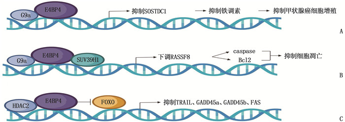
腺病毒E4启动子结合蛋白4(E4BP4)是一种含有碱性亮氨酸拉链的转录因子, 在乳腺癌、肺癌、甲状腺癌、结直肠癌等多种恶性肿瘤中高表达,参与肿瘤的发生发展、转移和侵袭,但关于E4BP4在肿瘤研究中的相关综述较少。本文检索并总结相关文献,概述了E4BP4在不同肿瘤发生发展中的作用,为深入研究E4BP4在肿瘤中的作用机制以及基于E4BP4分子治疗靶点的探究提供理论支撑。
-
关键词:
- 转录因子腺病毒E4启动子结合蛋白4 /
- 肿瘤 /
- 分子机制 /
- 治疗靶点 /
- 研究进展
Abstract:E4 promoter-binding protein 4 (E4BP4) is a transcription factor containing alkaline leucine zips, which is highly expressed in breast cancer, lung cancer, thyroid cancer, colorectal cancer and other malignant tumors, and is involved in the occurrence, development, metastasis and invasion of tumors. However, there are few reviews on E4BP4 in tumor research. In this paper, relevant literature was searched and summarized, and the role of E4BP4 in the occurrence and development of different tumors was summarized, so as to provide theoretical support for further study of the mechanism of action of E4BP4 in tumors and exploration of therapeutic targets based on E4BP4 molecules.
-
全膝关节置换术(TKA)是治疗终末期骨关节炎的重要方法,可有效改善关节活动度,矫正畸形,恢复关节功能,提高患者的生活质量[1]。随着骨性关节炎发病率的不断提高, TKA的临床开展率也不断上升。术后发热是TKA临床治疗过程中的常见症状,可分为感染性和非感染性,目前针对非感染性发热(NIF)的研究相对较少。与感染性发热不同的是, NIF无病原微生物感染的确切证据,但临床发生率更高,与手术、麻醉创伤以及机体炎症、应激反应有关[2-3], 往往会导致严重疼痛、伤口愈合延迟、住院时间延长和康复需求增加[4]。目前, NIF的确切机制和危险因素尚无定论,可影响NIF的早期诊断和干预,对患者术后康复造成不利影响。列线图模型可将重要的危险因素可视化,更加方便临床医护人员对患者进行评估。本研究探讨TKA患者术后7 d内NIF的影响因素并构建列线图预测模型,以期为临床早期诊断NIF提供一款简洁的量化工具,现报告如下。
1. 资料与方法
1.1 一般资料
采用回顾性队列研究方法,选取2017年1月—2021年7月在淮北市人民医院行单侧TKA治疗的201例膝骨关节炎患者作为研究对象,根据术后7 d内是否发生NIF将患者分为NIF组57例和无NIF组144例。纳入标准: ①年龄>18岁者; ②符合膝骨关节炎诊断标准[5]和TKA手术指征[6], 顺利完成手术且康复出院者; ③术后胸片、血尿、关节腔穿刺等检查未见明显感染征象者; ④临床资料完整者。排除标准: ①合并类风湿关节炎、骨肿瘤、骨外伤、痛风性关节炎等骨关节疾病者; ②近期有大手术、创伤史者; ③近期有重症感染、肝肾功能障碍、输血史者; ④围术期发生严重并发症(心肌梗死、中风、血管内凝血障碍、休克、肺栓塞等)者。
术后第2~7天,护士使用红外鼓膜温度计测量患者体温,每天至少测量4次(6: 00、10: 00、14: 00、18: 00)。NIF定义为术后7 d内任意时点体温≥38.0 ℃, 且胸片、血尿、关节腔穿刺等检查未见明显感染征象[7]。
1.2 方法
通过医院电子病历系统和护理记录单收集患者的临床资料,主要包括性别、年龄、基础疾病、手术时间、术中失血量、术后引流量、输血次数、抗生素使用时间和住院时间。所有患者的手术和术后护理均由经验丰富的手术团队和护理团队完成,术后采用头孢唑啉钠(1 g)预防感染, 2次/d, 持续48 h。术后48~72 h或日引流液量<100 mL时拔除引流管,术后根据患者康复情况开展功能锻炼。
1.3 统计学分析
采用SPSS 23.0统计学软件进行数据处理,正态分布的计量资料以(x±s)表示, 2组间比较采用独立样本t检验; 非正态分布的计量资料以[M(P25, P75)]表示,采用Mann-Whitney U检验; 计数资料以[n(%)]表示,比较采用χ2检验。采用LASSO回归模型筛选非零特征的变量,并采用多因素Logistic回归分析筛选影响因素,纳入标准α=0.10, 剔除标准β=0.05, 以逐步后退法进行筛选; 应用R软件和rms程序包建立列线图模型; 绘制受试者工作特征(ROC)曲线,计算列线图模型预测NIF的曲线下面积(AUC), 评估模型的区分度; 绘制Calibration校正曲线,评价模型的一致性; 通过决策曲线分析(DCA)确定列线图模型的临床有效性。检验水准为双侧α=0.05, 以P<0.05为差异有统计学意义。
2. 结果
2.1 NIF的单因素分析
NIF组术中失血量、术后引流量、输血者均多于无NIF组,手术时间、抗生素应用时间和住院时间均长于无NIF组,差异有统计学意义(P<0.05), 见表 1。
表 1 NIF的单因素分析(x±s)[M(P25, P75)][n(%)]指标 无NIF组(n=144) NIF组(n=57) Z/t/χ2 P 性别 男 28(19.4) 14(24.6) 0.314 0.575 女 116(80.6) 43(75.4) 年龄/岁 65.6±5.3 67.8±5.9 0.659 0.324 高血压 52(36.1) 18(31.6) 0.370 0.543 糖尿病 29(20.1) 7(12.3) 1.715 0.190 手术时间/min 177.9(126.5, 223.6) 212.5(165.4, 265.8) 2.063 <0.001 术前体温/℃ 36.8±0.4 36.7±0.3 0.235 0.867 术中失血量/mL 280.3(140.2, 420.3) 385.3(250.5, 530.6) 2.659 <0.001 术后引流量/mL 160.8(90.5, 320.8) 280.6(110.7, 450.9) 3.002 <0.001 输血 9(6.3) 13(22.8) 11.485 <0.001 抗生素使用时间/d 4.6(3.5, 7.5) 9.5(5.5, 15.5) 1.958 <0.001 住院时间/d 8.2±2.3 14.6±3.5 5.659 <0.001 2.2 NIF影响因素的LASSO回归分析
将单因素分析中差异有统计学意义的指标作为自变量,纳入LASSO回归模型,共筛选出4个具有非零特征的变量,即术中失血量、术后引流量、输血和手术时间,见图 1。
![]() 图 1 NIF影响因素的LASSO回归分析A: 利用最小标准值和10倍交叉验证识别LASSO模型中的λ; B: 显示特征的套索系数剖面
图 1 NIF影响因素的LASSO回归分析A: 利用最小标准值和10倍交叉验证识别LASSO模型中的λ; B: 显示特征的套索系数剖面
(使用10倍交叉验证在选定值处绘制垂直线,并利用最小标准值和最小标准值的1个标准误获得最佳值)。2.3 NIF影响因素的多因素Logistic回归分析
多因素Logistic回归分析显示,术中失血量、术后引流量、输血和手术时间是TKA患者术后7 d内NIF的独立影响因素(P<0.001), 见表 2。
表 2 NIF影响因素的多因素Logistic回归分析因素 β Wald P OR 95%CI 术中失血量 1.325 10.524 <0.001 3.652 2.856~3.958 术后引流量 1.002 8.623 <0.001 2.857 2.242~3.234 输血 1.712 16.596 <0.001 4.001 3.562~4.659 手术时间 0.758 5.965 <0.001 1.859 1.326~2.525 2.4 NIF的列线图模型
基于多因素Logistic回归分析筛选出的影响因素及其对应权重(β值),应用R软件构建列线图模型,见图 2。若1例患者术中失血量为400 mL(对应分值22分),术后引流量为400 mL(对应分值20分),输血(对应分值60分),手术时间为200 min(对应分值10分),则各项分值相加的总分为112分,对应风险为0.93, 提示其发生NIF的概率较高。
2.5 列线图模型预测NIF的内部验证结果
ROC曲线显示,列线图模型预测NIF的AUC为0.865(95%CI为0.799~0.901), 提示该模型的区分度和预测效能较好,见图 3。Calibration校正曲线显示,该模型的一致性较好,见图 4。DCA显示, NIF发生的风险阈值超过8%时,列线图模型的临床价值最大,见图 5。
3. 讨论
临床研究[8]发现, TKA后NIF十分常见,发生时间多为术后1~10 d, 高峰期为术后7 d内,故本研究主要统计术后7 d内NIF发生情况。NIF的主要临床表现为无明显感染迹象的体温升高,抗生素应用效果不佳,发热持续时间较长,可影响手术疗效,造成术后康复质量不佳[9]。研究[10-11]发现,组织损伤、药物、输血反应、贫血和深静脉血栓形成等非感染性因素可以刺激机体释放大量炎症介质,导致体温升高,但各种因素的主次关系仍未阐明,为临床早期干预增加了难度。目前,临床尚无公认的可靠指标来预测NIF的发生,故本研究基于NIF的危险因素构建列线图模型,以期为临床早期诊断NIF提供有效的工具。
本研究显示, TKA患者术后7 d内NIF发生率为28.4%(57/201), 与相关研究[12-13]报道的发生率差异较大,这可能与纳入样本量及患者病情特征不同有关。针对术后发热,临床首先需要通过相关指标结果排除感染性原因,但往往需要抽血、关节腔抽液、细菌培养以及影像学检查等,会增加患者痛苦和医疗费用,而大多数患者检测结果为阴性。本研究结合实际情况比较分析了2组患者术中失血量、输血、术后引流量等容易监测的可影响液体平衡和血容量的因素,这些因素与机体的体液平衡显著相关,而体液平衡是维持体温调节能力的关键因素[14-15]。本研究结果显示, NIF组术中失血量、术后引流量、输血者均多于无NIF组,手术时间、抗生素应用时间和住院时间均长于无NIF组,差异有统计学意义(P<0.05)。本研究利用LASSO回归分析筛选非零特征的变量(降低变量间的相关性,突出变量的独立性),并进行多因素Logistic回归分析,结果显示,术中失血量、术后引流量、输血和手术时间均为TKA患者术后7 d内NIF的独立影响因素。术中失血量和术后引流量是影响循环血容量稳定的重要原因,液体丢失可导致体温调节不稳定[16]。减少异体输血有助于预防发热并发症,尤其是在大型骨科手术中[17]。另有研究[18]显示,手术后排出的液体量与局部血管损伤、伤口渗出和金属刺激有关。手术损伤区域的出血和渗出会刺激局部组织发生炎症反应,使机体释放大量肿瘤坏死因子、白细胞介素以及其他细胞因子,促进内源性热源的产生,导致患者体温升高[19-20]。
本研究应用R软件构建列线图模型(基于NIF发生的独立影响因素),并进行多维度验证。ROC曲线显示,该列线图模型预测NIF的AUC为0.865, 提示模型的区分度和预测效能较好; Calibration校正曲线显示,该模型的一致性较好; DCA显示, NIF发生的风险阈值超过8%时,该模型的临床价值最大。本研究存在一定局限性,例如样本量和观察时间有限,数据来源于单中心回顾性病例分析结果,且列线图模型仅进行了内部验证,缺乏外部验证数据,故其准确性和推广价值有待进一步深入研究后加以验证。
综上所述, TKA患者术后7 d内NIF发生率较高,术中失血量、术后引流量、输血和手术时间是NIF发生的独立影响因素,基于影响因素构建的列线图模型可视化效果较好,预测NIF发生的效能较高。
-
[1] COWELL I G, SKINNER A, HURST H C. Transcriptional repression by a novel member of the bZIP family of transcription factors[J]. Mol Cell Biol, 1992, 12(7): 3070-3077.
[2] ZHANG W, ZHANG J, KORNUC M, et al. Molecular cloning and characterization of NF-IL3A, a transcriptional activator of the human interleukin-3 promoter[J]. Mol Cell Biol, 1995, 15(11): 6055-6063. doi: 10.1128/MCB.15.11.6055
[3] HURST H C. Transcription factors 1: bZIP proteins[J]. Protein Profile, 1995, 2(2): 101-168.
[4] WANG Y H, KUANG Z, YU X F, et al. The intestinal microbiota regulates body composition through NFIL3 and the circadian clock[J]. Science, 2017, 357(6354): 912-916. doi: 10.1126/science.aan0677
[5] VELMURUGAN B K, CHANG R L, MARTHANDAM ASOKAN S, et al. A minireview of E4BP4/NFIL3 in heart failure[J]. J Cell Physiol, 2018, 233(11): 8458-8466. doi: 10.1002/jcp.26790
[6] LI D, WANG Y D, YANG M X, et al. mTORC1 and mTORC2 coordinate early NK cell development by differentially inducing E4BP4 and T-bet[J]. Cell Death Differ, 2021, 28(6): 1900-1909. doi: 10.1038/s41418-020-00715-6
[7] HIRAI T, TANAKA K, TOGARI A. Β-adrenergic receptor signaling regulates Ptgs2 by driving circadian gene expression in osteoblasts[J]. J Cell Sci, 2014, 127(Pt 17): 3711-3719.
[8] YIN J H, ZHANG J, LU Q J. The role of basic leucine zipper transcription factor E4BP4 in the immune system and immune-mediated diseases[J]. Clin Immunol, 2017, 180: 5-10. doi: 10.1016/j.clim.2017.03.013
[9] 王健, 黄哲平, 杨增明, 等. 腺病毒E4启动子结合蛋白-4(E4BP4)基因在小鼠胚胎着床期间子宫组织中的表达[J]. 分子细胞生物学报, 2006, 39(2): 123-131. https://www.cnki.com.cn/Article/CJFDTOTAL-SWSB200602006.htm [10] KENIRY M, PIRES M M, MENSE S, et al. Survival factor NFIL3 restricts FOXO-induced gene expression in cancer[J]. Genes Dev, 2013, 27(8): 916-927. doi: 10.1101/gad.214049.113
[11] QI J J, YU Y, AKILLI ÖZTVRK Ö, et al. New Wnt/β-catenin target genes promote experimental metastasis and migration of colorectal cancer cells through different signals[J]. Gut, 2016, 65(10): 1690-1701. doi: 10.1136/gutjnl-2014-307900
[12] KURIBARA R, KINOSHITA T, MIYAJIMA A, et al. Two distinct interleukin-3-mediated signal pathways, Ras-NFIL3 (E4BP4) and Bcl-xL, regulate the survival of murine pro-B lymphocytes[J]. Mol Cell Biol, 1999, 19(4): 2754-2762. doi: 10.1128/MCB.19.4.2754
[13] ZHU C, SAKUISHI K, XIAO S, et al. An IL-27/NFIL3 signalling axis drives Tim-3 and IL-10 expression and T-cell dysfunction[J]. Nat Commun, 2015, 6: 6072. doi: 10.1038/ncomms7072
[14] RHODES D R, KALYANA-SUNDARAM S, MAHAVISNO V, et al. Oncomine 3. 0: genes, pathways, and networks in a collection of 18, 000 cancer gene expression profiles[J]. Neoplasia, 2007, 9(2): 166-180.
[15] LIN S C, LIN C H, SHIH N C, et al. Cellular prion protein transcriptionally regulated by NFIL3 enhances lung cancer cell lamellipodium formation and migration through JNK signaling[J]. Oncogene, 2020, 39(2): 385-398. doi: 10.1038/s41388-019-0994-0
[16] 陆宁宁, 邾萍, 马珠月, 等. 乳腺癌患者一级亲属乳腺癌早期筛查的研究进展[J]. 实用临床医药杂志, 2021, 25(22): 129-132. doi: 10.7619/jcmp.20212780 [17] LIU L, WU S S, YANG Y, et al. SOSTDC1 is down-regulated in non-small cell lung cancer and contributes to cancer cell proliferation[J]. Cell Biosci, 2016, 6: 24. doi: 10.1186/s13578-016-0091-9
[18] GOPAL G, RAJA U M, SHIRLEY S, et al. SOSTDC1 down-regulation of expression involves CpG methylation and is a potential prognostic marker in gastric cancer[J]. Cancer Genet, 2013, 206(5): 174-182. doi: 10.1016/j.cancergen.2013.04.005
[19] RAWAT A, GOPISETTY G, THANGARAJAN R. E4BP4 is a repressor of epigenetically regulated SOSTDC1 expression in breast cancer cells[J]. Cell Oncol (Dordr), 2014, 37(6): 409-419.
[20] KARTHIK I P, DESAI P, SUKUMAR S, et al. E4BP4/NFIL3 modulates the epigenetically repressed RAS effector RASSF8 function through histone methyltransferases[J]. J Biol Chem, 2018, 293(15): 5624-5635. doi: 10.1074/jbc.RA117.000623
[21] 周树伟, 苏蓓蓓, 冯跃庆, 等. 分化型甲状腺癌患者血清基质金属蛋白酶-13表达水平与肺转移的相关性研究[J]. 实用临床医药杂志, 2021, 25(1): 77-80. doi: 10.7619/jcmp.20200593 [22] ZHOU Q Y, CHEN J, FENG J L, et al. E4BP4 promotes thyroid cancer proliferation by modulating iron homeostasis through repression of hepcidin[J]. Cell Death Dis, 2018, 9(10): 987. doi: 10.1038/s41419-018-1001-3
[23] ZHOU Q Y, CHEN J, FENG J L, et al. SOSTDC1 inhibits follicular thyroid cancer cell proliferation, migration, and EMT via suppressing PI3K/Akt and MAPK/Erk signaling pathways[J]. Mol Cell Biochem, 2017, 435(1/2): 87-95.
[24] CHA S, SIN M J, KIM M J, et al. Involvement of cellular prion protein in invasion and metastasis of lung cancer by inducing treg cell development[J]. Biomolecules, 2021, 11(2): 285. doi: 10.3390/biom11020285
[25] FRITZMANN J, MORKEL M, BESSER D, et al. A colorectal cancer expression profile that includes transforming growth factor beta inhibitor BAMBI predicts metastatic potential[J]. Gastroenterology, 2009, 137(1): 165-175. doi: 10.1053/j.gastro.2009.03.041
[26] XU J X, XU G P, ZHANG T X, et al. NFIL3 acts as a nuclear factor to increase osteosarcoma progression[J]. Biomed Res Int, 2019, 2019: 4068521.
[27] LURAIN J R. Pharmacotherapy of gestational trophoblastic disease[J]. Expert Opin Pharmacother, 2003, 4(11): 2005-2017. doi: 10.1517/14656566.4.11.2005
[28] PENG Z, ZHANG C, ZHOU W J, et al. The STAT3/NFIL3 signaling axis-mediated chemotherapy resistance is reversed by Raddeanin A via inducing apoptosis in choriocarcinoma cells[J]. J Cell Physiol, 2018, 233(7): 5370-5382. doi: 10.1002/jcp.26362
[29] BEACH J A, NARY L J, HIRAKAWA Y, et al. E4BP4 facilitates glucocorticoid-evoked apoptosis of human leukemic CEM cells via upregulation of Bim[J]. J Mol Signal, 2011, 6(1): 13.
[30] BEACH J A, NARY L J, HOVANESSIAN R, et al. Correlation of glucocorticoid-mediated E4BP4 upregulation with altered expression of pro-and anti-apoptotic genes in CEM human lymphoblastic leukemia cells[J]. Biochem Biophys Res Commun, 2014, 451(3): 382-388. doi: 10.1016/j.bbrc.2014.07.103
[31] PRICEMAN S J, KIRZNER J D, NARY L J, et al. Calcium-dependent upregulation of E4BP4 expression correlates with glucocorticoid-evoked apoptosis of human leukemic CEM cells[J]. Biochem Biophys Res Commun, 2006, 344(2): 491-499. doi: 10.1016/j.bbrc.2006.03.169
[32] NISHIMURA Y, TANAKA T. Calcium-dependent activation of nuclear factor regulated by interleukin 3/adenovirus E4 promoter-binding protein gene expression by calcineurin/nuclear factor of activated T cells and calcium/calmodulin-dependent protein kinase signaling[J]. J Biol Chem, 2001, 276(23): 19921-19928. doi: 10.1074/jbc.M010332200
[33] DORN D C, KOU C A, PNG K J, et al. The effect of cantharidins on leukemic stem cells[J]. Int J Cancer, 2009, 124(9): 2186-2199. doi: 10.1002/ijc.24157
[34] YEUNG J, O'SULLIVAN E, HUBANK M, et al. E4BP4 expression is regulated by the t(17; 19)-associated oncoprotein E2A-HLF in pro-B cells[J]. Br J Haematol, 2004, 125(5): 560-567. doi: 10.1111/j.1365-2141.2004.04953.x
[35] WU S J, LI J Y, CAO M S, et al. A novel integrated gene coexpression analysis approach reveals a prognostic three-transcription-factor signature for glioma molecular subtypes[J]. BMC Syst Biol, 2016, 10(Suppl 3): 71.
[36] KENIRY M, DEARTH R K, PERSANS M, et al. New frontiers for the NFIL3 bZIP transcription factor in cancer, metabolism and beyond[J]. Discoveries (Craiova), 2014, 2(2): e15.
[37] UNOKI M, NAKAMURA Y. Growth-suppressive effects of BPOZ and EGR2, two genes involved in the PTEN signaling pathway[J]. Oncogene, 2001, 20(33): 4457-4465.
[38] SADOWSKI S M, PUSZTASZERI M, BRULHART-MEYNET M C, et al. Identification of differential transcriptional patterns in primary and secondary hyperparathyroidism[J]. J Clin Endocrinol Metab, 2018, 103(6): 2189-2198.
 下载:
下载:


计量
- 文章访问数: 188
- HTML全文浏览量: 104
- PDF下载量: 15
 苏公网安备 32100302010246号