| Citation: |
CAO Na, HUANG Boqi, LIU Zhimin, WANG Dong. Establish of a simple predictive model based on CT imaging histology in the differential diagnosis of extra-small renal cell carcinoma with rich blood supply and angiomyolipoma with minimal fat[J]. Journal of Clinical Medicine in Practice, 2023, 27(10): 6-11. DOI: 10.7619/jcmp.20230492

|
To explore the differential value of a simple prediction model based on CT imaging histology in the diagnosis of ultra-small renal cell carcinoma (usRCC) with rich blood supply and angiomyolipoma with minimal fat (mfAML).
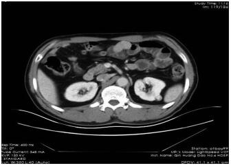
The clinical data of 71 patients with ultra-small renal tumor (diameter ≤2 cm) with rich blood supply were collected. According to the postoperative pathological types, they were divided into usRCC group (n=33) and mfAML group (n=38). Clinical data, CT imaging manifestations, and related CT quantitative parameters were compared, independent influencing factors with differential significance for usRCC and mfAML were screened using binary Logistic regression, and a simple predictive model based on CT imaging histology was constructed. Receiver operating characteristic (ROC) curves were drawn to evaluate differential value of relevant CT quantitative parameters and predictive model for usRCC and mfAML.
The proportion of cystic necrosis, pseudocapsule sign and parenchymal phase heterogeneous enhancement in the usRCC group was higher than that in the mfAML group (P < 0.05). The CT value of cortical phase, enhanced CT value of cortical phase and parenchymal phase in the usRCC group were also significantly higher than those in the mfAML group (P < 0.05). The areas under the curve (AUCs) of differential diagnosis of usRCC and mfAML by CT value of cortical phase, enhanced CT value of cortical phase and parenchymal phase were 0.702, 0.718 and 0.803, respectively. Cystic necrosis (OR=2.537; 95% CI, 1.125 to 4.358), parenchymal enhancement uniformity (OR=3.872; 95% CI, 1.327 to 7.259), and parenchymal net enhancement CT value (OR=3.593; 95% CI, 1.290 to 7.518) were independent influencing factors for differentiated diagnosis of usRCC and mfAML (P < 0.05), thus a simple prediction model was constructed based on the three CT image omics variables. The ROC curve showed that the AUC of the model for differential diagnosis of usRCC and mfAML was 0.890 (95% CI, 0.804 to 0.976), the sensitivity was 87.888, and the specificity was 76.32%.
The simple prediction model based on CT imaging histology has a good value in differential diagnosis of usRCC and mfAML, and provides an important reference for clinical diagnosis and treatment of small renal tumors.
| [1] |
王禹, 董潇, 孔垂泽, 等. 不同病理类型肾肿瘤的影像学特点和病理学特点分析[J]. 中华泌尿外科杂志, 2019, 40(5): 374-379. https://cdmd.cnki.com.cn/Article/CDMD-10159-1021870603.htm
|
| [2] |
程波, 邹菊, 向碧兰, 等. LncRNA在肾癌诊治及预后方面的研究进展[J]. 西南医科大学学报, 2022, 45(3): 272-276. https://www.cnki.com.cn/Article/CJFDTOTAL-LXYB202203018.htm
|
| [3] |
张丽青, 韩志江. 最大径≤3 cm的乏脂肪肾脏血管平滑肌脂肪瘤与肾透明细胞癌的MRI鉴别[J]. 临床放射学杂志, 2019, 38(1): 132-135. https://www.cnki.com.cn/Article/CJFDTOTAL-LCFS201901031.htm
|
| [4] |
崔梦秋, 王海屹, 许伟, 等. 最大径≤4 cm肾脏血管平滑肌脂肪瘤的MRI征象分析[J]. 中华放射学杂志, 2022, 56(5): 549-555.
|
| [5] |
YE J, XU Q, WANG S A, et al. Differentiation between fat-poor angiomyolipoma and clear cell renal cell carcinoma: qualitative and quantitative analysis using arterial spin labeling MR imaging[J]. Abdom Radiol, 2020, 45(2): 512-519. doi: 10.1007/s00261-019-02303-w
|
| [6] |
程劲松, 韩津梁. 应用MRI鉴别小肾癌与乏脂型血管平滑肌脂肪瘤的研究[J]. 中国实验诊断学, 2018, 22(3): 478-480. https://www.cnki.com.cn/Article/CJFDTOTAL-ZSZD201803032.htm
|
| [7] |
李庆芬, 赵丽雅, 苏博, 等. 肾肿瘤的彩色多普勒超声诊断价值探讨[J]. 保健医学研究与实践, 2012, 9(4): 37-39. https://www.cnki.com.cn/Article/CJFDTOTAL-GXBJ201204014.htm
|
| [8] |
孙翌峰, 李建瑞, 陈子健, 等. 多排螺旋CT对不同亚型肾癌、肾血管平滑肌脂肪瘤和肾嗜酸细胞腺瘤的诊断价值[J]. 医学影像学杂志, 2018, 28(5): 794-799. https://www.cnki.com.cn/Article/CJFDTOTAL-XYXZ201805030.htm
|
| [9] |
沈剑, 杨印辉, 孙颖浩, 等. 疑似肾癌的18例肾良性肿瘤诊治分析[J]. 实用临床医药杂志, 2012, 16(5): 107-109. doi: 10.3969/j.issn.1672-2353.2012.05.038
|
| [10] |
ZHAO X F, JHALA N, KHURANA J. Evaluation of radiology imaging in diagnosing clear cell renal cell carcinoma and angiomyolipoma[J]. Am J Clin Pathol, 2018, 150(suppl_1): S27. http://www.researchgate.net/publication/327812133_Evaluation_of_Radiology_Imaging_in_Diagnosing_Clear_Cell_Renal_Cell_Carcinoma_and_Angiomyolipoma
|
| [11] |
LIANG X H, XUE C Q, HUANG X Y, et al. Value of energy spectrum CT parameters in the differential diagnosis of high-grade clear cell renal cell carcinoma and type Ⅱ papillary renal cell carcinoma[J]. Acta Radiol, 2022, 63(4): 545-552. http://pubmed.ncbi.nlm.nih.gov/33779302/
|
| [12] |
王思凯, 李鸣瑶, 梁卡丽, 等. 多排螺旋CT诊断乏脂性肾血管平滑肌脂肪瘤与肾癌的对照研究[J]. 中国医学计算机成像杂志, 2018, 24(3): 215-218. https://www.cnki.com.cn/Article/CJFDTOTAL-YJTY201803007.htm
|
| [13] |
向玲玲, 吴晶涛, 郜言坤, 等. 增强CT影像组学鉴别小肾癌与乏脂肪肾血管平滑肌脂肪瘤的价值[J]. 肿瘤影像学, 2021, 30(3): 185-190. https://www.cnki.com.cn/Article/CJFDTOTAL-YXYX202103008.htm
|
| [14] |
LEE H S, HONG H, KIM J, et al. Deep feature classification of angiomyolipoma without visible fat and renal cell carcinoma in abdominal contrast-enhanced CT images with texture image patches and hand-crafted feature concatenation[J]. Med Phys, 2018, 45(4): 1550-1561. doi: 10.1002/mp.12828/pdf
|
| [15] |
高原, 蔡晓娟, 陆建东. 增强CT鉴别诊断乏脂性肾血管平滑肌脂肪瘤与肾癌的影像特征及临床价值[J]. 癌症进展, 2018, 16(10): 1231-1233, 1237. https://www.cnki.com.cn/Article/CJFDTOTAL-AZJZ201810010.htm
|
| [16] |
黄裕存, 黄胜福, 陆少范, 等. CT形态学特征鉴别乏脂质血管平滑肌脂肪瘤和肾细胞癌[J]. 中国CT和MRI杂志, 2020, 18(6): 107-109, 153. https://www.cnki.com.cn/Article/CJFDTOTAL-CTMR202006034.htm
|
| [17] |
罗敏, 蔡文超, 张玮, 等. 肾上皮样血管平滑肌脂肪瘤(长径≤3 cm)的影像诊断[J]. 放射学实践, 2018, 33(12): 1295-1301. https://www.cnki.com.cn/Article/CJFDTOTAL-FSXS201812017.htm
|
| [18] |
王旭, 宋歌, 王宗平, 等. 早期富血供超小肾癌与肾乏脂肪血管平滑肌脂肪瘤的CT鉴别诊断[J]. 中华全科医学, 2020, 18(6): 989-993, 1017. https://www.cnki.com.cn/Article/CJFDTOTAL-SYQY202006028.htm
|
| [19] |
赵小芳, 于志鹏, 赵长秀, 等. 乏脂性肾血管平滑肌脂肪瘤与小肾癌的CT鉴别诊断价值[J]. 现代医用影像学, 2021, 30(10): 1838-1841. https://www.cnki.com.cn/Article/CJFDTOTAL-XDYY202110012.htm
|
| [20] |
CUI E M, LIN F, LI Q, et al. Differentiation of renal angiomyolipoma without visible fat from renal cell carcinoma by machine learning based on whole-tumor computed tomography texture features[J]. Acta Radiol, 2019, 60(11): 1543-1552.
|
| [21] |
黄忠江, 姜增誉, 李健丁, 等. 基于增强CT影像组学联合机器学习鉴别均质性肾透明细胞癌与肾乏脂肪血管平滑肌脂肪瘤[J]. 实用医学杂志, 2021, 37(17): 2266-2270. https://www.cnki.com.cn/Article/CJFDTOTAL-SYYZ202117020.htm
|
| [22] |
崔志勇, 崔二峰, 王刚, 等. 应用MSCT鉴别诊断AML、非透明细胞肾癌的效果分析[J]. 中国CT和MRI杂志, 2021, 19(6): 113-115. https://www.cnki.com.cn/Article/CJFDTOTAL-CTMR202106037.htm
|
| 1. |
李娜,包娜日素,王丽斯,李敏,张生茂. 不同通气模式对腹腔镜结直肠癌手术患者呼吸力学、血流动力学和生化代谢的影响. 中国医师进修杂志. 2023(05): 449-454 .

| |
| 2. |
唐婷,张庆. 压力控制与肺保护性容量控制通气在俯卧位腰椎手术中的应用比较. 中国现代医药杂志. 2021(04): 38-41 .
|
© 2020 《实用临床医药杂志》编辑部
Address: 江苏省扬州市江阳中路136号,扬州大学江阳路北校区14号楼201室China Pos: 225009Tel: 0514-87978917、87978989、87978807
Supported by:
Beijing Renhe Information Technology Co., Ltd.

 苏公网安备 32100302010246号
 DownLoad: