| Citation: |
YUE Mengchao, LIU Yang, XIE Ying. Diagnostic value of dynamic contrast-enhanced magnetic resonance imaging and signal intensity-time curve in benign and malignant prostate lesions[J]. Journal of Clinical Medicine in Practice, 2023, 27(15): 82-85. DOI: 10.7619/jcmp.20231559

|
To explore the diagnostic value of dynamic contrast-enhanced magnetic resonance imaging (DCE-MRI) and signal intensity-time (SI-T) curve in benign and malignant prostate lesions.
A total of 102 patients with prostate disease were selected as the study objects, and were divided into benign group (65 cases) and malignant group (37 cases). Receiver operating characteristic (ROC) curve was used to analyze the diagnostic value of DCE-MRI, SI-T alone and their combined detection in benign and malignant prostate lesions.
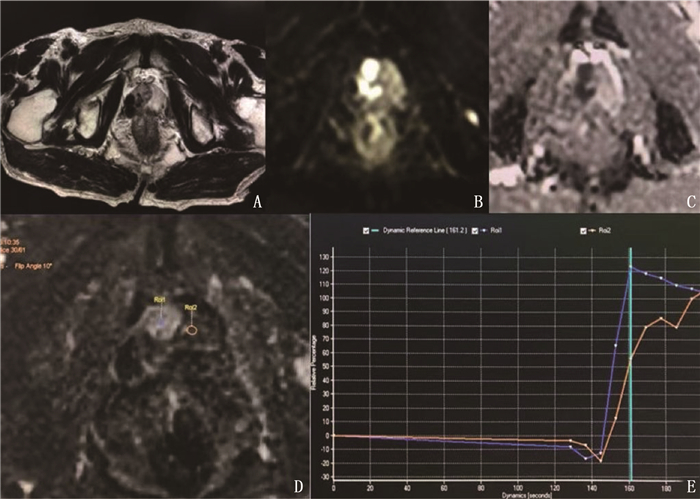
The volume transfer constant (Ktrans), rate constant (Kep) and extravascular extracellular space volume fraction (Ve) of patients in the malignant group were higher than those in the benign group (P < 0.05). The proportion of fast growth and slow decline of SI-T curve in the malignant group was significantly higher than that in the benign group, and the proportion of continuous slow rise and slow rise platform type was significantly lower than that in the benign group (P < 0.05). The peak time of the malignant group was significantly shorter than that of the benign group, and the degree and rate of reinforcement were significantly higher than those of the benign group (P < 0.05). The area under the curve (AUC) of the combined detection of DCE-MRI or SI-T curve parameter in the diagnosis of benign and malignant prostate lesions was greater than that of the single detection of DCE-MRI or SI-T curve parameter (P < 0.05).
The combined detection of DCE-MRI and SI-T curve has good diagnostic value in diagnosis of benign and malignant prostate lesions.
| [1] |
CHANG Y X, DENG Q, GUAN Z F, et al. miR-1273g-3p promotes malignant progression and has prognostic implications in prostate cancer[J]. Mol Biotechnol, 2022, 64(1): 17-24. doi: 10.1007/s12033-021-00384-x
|
| [2] |
余英芳, 胡海霞, 王小义, 等. 动态对比增强MRI在肺良恶性结节(≤2 cm)的诊断价值[J]. 中国CT和MRI杂志, 2022, 20(11): 64-66. https://www.cnki.com.cn/Article/CJFDTOTAL-CTMR202211024.htm
|
| [3] |
刘晓东, 方习奇, 唐桑, 等. 动态对比增强MRI联合表观扩散系数值对前列腺中央区癌的诊断价值[J]. 实用放射学杂志, 2020, 36(4): 599-602.
|
| [4] |
中国抗癌协会泌尿男生殖系肿瘤专业委员会微创学组. 中国前列腺癌外科治疗专家共识[J]. 浙江医学, 2018, 40(3): 217-220. https://www.cnki.com.cn/Article/CJFDTOTAL-ZGAZ202212013.htm
|
| [5] |
ALLARAKHA A, GAO Y, JIANG H, et al. Predictive ability of DWI/ADC and DCE-MRI kinetic parameters in differentiating benign from malignant breast lesions and in building a prediction model[J]. Discov Med, 2019, 27(148): 139-152.
|
| [6] |
NAKANISHI K, TANAKA J, NAKAYA Y, et al. Whole-body MRI: detecting bone metastases from prostate cancer[J]. Jpn J Radiol, 2022, 40(3): 229-244. doi: 10.1007/s11604-021-01205-6
|
| [7] |
孙文杰, 王欣, 刘玲, 等. 3. 0T磁共振多参数成像及动态增强扫描对前列腺癌的诊断价值[J]. 中国CT和MRI杂志, 2022, 20(1): 138-141. https://www.cnki.com.cn/Article/CJFDTOTAL-CTMR202201043.htm
|
| [8] |
任义财, 周利平. DCE-MRI、3D1H-MRS联合对前列腺良恶性病变的诊断价值[J]. 中国CT和MRI杂志, 2020, 18(4): 107-109, 113. https://www.cnki.com.cn/Article/CJFDTOTAL-CTMR202004032.htm
|
| [9] |
杜元元, 李玉智, 马蔚, 等. DWI、1H-MRS及DCE-MR对前列腺良恶性病变的诊断价值[J]. 海南医学, 2022, 33(3): 352-356. https://www.cnki.com.cn/Article/CJFDTOTAL-HAIN202203020.htm
|
| [10] |
贾海云, 王佳强, 周晓莹, 等. 剪切波弹性成像联合多参数MRI对临床显著性前列腺癌的诊断价值[J]. 临床超声医学杂志, 2022, 24(8): 603-607. https://www.cnki.com.cn/Article/CJFDTOTAL-LCCY202208008.htm
|
| [11] |
李鹏, 黄英, 李艳, 等. DWI和DCE-MRI鉴别诊断良恶性前列腺外周带T2WI局灶性低信号病变[J]. 中国介入影像与治疗学, 2021, 18(3): 156-160. https://www.cnki.com.cn/Article/CJFDTOTAL-JRYX202103011.htm
|
| [12] |
刘郭坤, 李健斐, 刘艳超, 等. IVIM-DWI与DCE-MRI在前列腺疾病诊断中的联合应用观察[J]. 山东医药, 2020, 60(3): 75-77. https://www.cnki.com.cn/Article/CJFDTOTAL-SDYY202003023.htm
|
| [13] |
STABILE A, BARLETTA F, MOTTERLE G, et al. Optimizing prostate-targeted biopsy schemes in men with multiple mpMRI visible lesions: should we target all suspicious areas Results of a two institution series[J]. Prostate Cancer Prostatic Dis, 2021, 24(4): 1137-1142. doi: 10.1038/s41391-021-00371-y
|
| [14] |
MONTORSI F, STABILE A, GANDAGLIA G, et al. Re: Marra etal. 'Transperineal freehand multiparametric MRI fusion targeted biopsies under local anaesthesia for prostate cancer diagnosis: a multicentre prospective study of 1014 cases'[J]. BJU Int, 2021, 128(4): 523.
|
| [15] |
刘凯, 罗红兰, 柯楠, 等. DCE-MRI和IVIM-DWI在前列腺癌病理分级和临床分期中的诊断价值分析[J]. 中国性科学, 2022, 31(7): 31-35. https://www.cnki.com.cn/Article/CJFDTOTAL-XKXZ202207009.htm
|
| [16] |
陈爽, 陈子健, 钟建锋, 等. 动态对比增强磁共振联合PSA诊断前列腺癌价值探讨[J]. 中国CT和MRI杂志, 2022, 20(1): 142-145. https://www.cnki.com.cn/Article/CJFDTOTAL-CTMR202201044.htm
|
| [17] |
GASSENMAIER S, AFAT S, NICKEL D, et al. Deep learning-accelerated T2-weighted imaging of the prostate: reduction of acquisition time and improvement of image quality[J]. Eur J Radiol, 2021, 137: 109600.
|
| [18] |
邰兆琴, 徐小虎, 许亚春, 等. 磁共振动态增强联合DWI与超声引导穿刺对照在前列腺病变诊断中的应用[J]. 中国CT和MRI杂志, 2021, 19(8): 113-116. https://www.cnki.com.cn/Article/CJFDTOTAL-CTMR202108037.htm
|
| [19] |
WEI C G, CHEN T, ZHANG Y Y, et al. Erratum to"Biparameteric prostate MRI and clinical indicators predict clinically significant prostate cancer in men with" gray zone "PSA levels"[J]. Eur J Radiol, 2020, 129: 109129.
|
| [20] |
李珍, 邹磊, 弥长虹, 等. 前列腺癌磁共振动态增强定量研究[J]. 中国CT和MRI杂志, 2021, 19(7): 126-127, 137. https://www.cnki.com.cn/Article/CJFDTOTAL-CTMR202107036.htm
|
| [21] |
PESAPANE F, STANDAERT C, VISSCHERE P D, et al. T-staging of prostate cancer: identification of useful signs to standardize detection of posterolateral extraprostatic extension on prostate MRI[J]. Clin Imaging, 2020, 59(1): 1-7.
|
| [22] |
陈莉, 黄道煌, 骆祥伟, 等. 3. 0T磁共振动态增强定量参数联合DWI诊断前列腺癌临床价值[J]. 医学影像学杂志, 2022, 32(6): 1073-1075. https://www.cnki.com.cn/Article/CJFDTOTAL-XYXZ202206043.htm
|
| [23] |
STONE L. Piloting hyperpolarized 13C-pyruvate MRI for imaging advanced prostate cancer[J]. Nat Rev Urol, 2020, 17(1): 7.
|
| [24] |
PARRA N A, LU H, LI Q, et al. Erratum: predicting clinically significant prostate cancer using DCE-MRI habitat descriptors[J]. Oncotarget, 2019, 10(21): 2113.
|
| [25] |
李盈盈, 张明博, 吴凡, 等. 经直肠多模态超声与多模态MRI检查对前列腺癌诊断价值的对照研究[J]. 中华泌尿外科杂志, 2020, 41(1): 19-25. https://cpfd.cnki.com.cn/Article/CPFDTOTAL-CSYX201810001237.htm
|
| [26] |
CHRISTOPHE C, MONTAGNE S, BOURRELIER S, et al. Prostate cancer local staging using biparametric MRI: assessment and comparison with multiparametric MRI[J]. Eur J Radiol, 2020, 132: 109350.
|
© 2020 《实用临床医药杂志》编辑部
Address: 江苏省扬州市江阳中路136号,扬州大学江阳路北校区14号楼201室China Pos: 225009Tel: 0514-87978917、87978989、87978807
Supported by:
Beijing Renhe Information Technology Co., Ltd.

 苏公网安备 32100302010246号
 DownLoad: