| Citation: |
CAO Juan, WANG Hongyi, WANG Lixin, CHEN Xiaoning, LIN Qiu, YANG Qinghong, CHEN Dongzhui, FU Yuxiu. Effects of electroacupuncture on hippocampal neuron autophagy and IGF-1/PI3K/Akt pathway in rats with hypoxic-ischemic brain damage[J]. Journal of Clinical Medicine in Practice, 2022, 26(14): 38-45. DOI: 10.7619/jcmp.20214746

|
To explore the effects of electroacupuncture on hippocampal neuron autophagy and insulin-like growth factor 1 (IGF-1)/phosphatidylinositol 3-kinase (PI3K)/protein kinase B (Akt) pathway in neonatal rats with hypoxic-ischemic brain damage (HIBD).
A total of 108 neonatal rats were randomly divided into sham operation group, model group, IGF-1 group (0.2 mg/kg), electroacupuncture group, electroacupuncture combined with LY294002 group (electroacupuncture combined with 0.3 mg/kg PI3K inhibitor), with 18 rats in each group. HIBD model of neonatal rats was established by ligation of left common carotid artery and hypoxia treatment for 2 hours (the success rate of modeling was 80%). The rats in each group were scored for neurological deficits at being awake after the operation and after electroacupuncture treatment; ELISA was used to detect the content of IGF-1 in brain tissues; hematoxylin-eosin (HE) staining was used to observe the pathological change of brain tissues; transmission electron microscope was used to observe the cell autophagy; immunofluorescence double-labeling method was used to detect the colocalization expression of autophagy markers and neuron-specific nuclear protein (NeuN); western blot method was used to detect the expression of PI3K/Akt pathway and autophagy-related proteins in hippocampus[PI3K, phosphorylated PI3K (p-PI3K), Akt, phosphorylated Akt (p-Akt), Beclin-1, microtubule-related protein light chain 3Ⅱ (LC3-Ⅱ), and microtubule-related protein light chain 3Ⅰ (LC3-Ⅰ)].
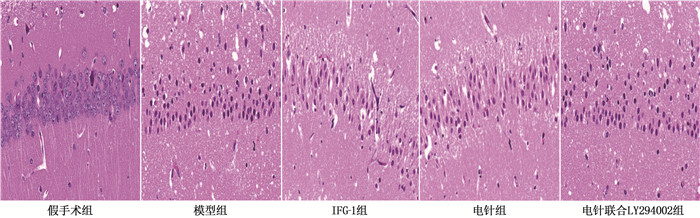
Compared with the sham operation group, the neuron damage in the hippocampus of the HIBD model group was aggravated, the neurological deficit score, the Beclin-1 and LC3-Ⅱ/LC3-I expression in the hippocampus were increased, while the IGF-1 content in brain tissues, p-PI3K/PI3K and p-Akt/Akt expression in hippocampus were reduced; compared with the model group, the neuron damage in the IGF-1 group and the electroacupuncture group was alleviated, the neurological deficit score, Beclin-1 and LC3-Ⅱ/LC3-Ⅰ, the number of autophagosomes and the LC3/NeuN co-expressed neurons were decreased or reduced, while the IGF-1 content in brain tissues, p-PI3K/PI3K and p-Akt/Akt expression were increased; the differences mentioned above were statistically significant (P<0.05). LY294002 was able to significantly reduce the level of IGF-1, weaken the activation of the IGF-1/PI3K/Akt pathway of hippocampus and the inhibition of hippocampal neuron autophagy by electroacupuncture.
Electroacupuncture may inhibit excessive autophagy of hippocampal neurons by activating the IGF-1/PI3K/Akt pathway, thereby alleviating HIBD in neonatal rats.
| [1] |
GRECO P, NENCINI G, PIVA I, et al. Pathophysiology of hypoxic-ischemic encephalopathy: a review of the past and a view on the future[J]. Acta Neurol Belg, 2020, 120(2): 277-288. doi: 10.1007/s13760-020-01308-3
|
| [2] |
YANG L J, ZHAO H H, CUI H. Treatment and new progress of neonatal hypoxic-ischemic brain damage[J]. Histol Histopathol, 2020, 35(9): 929-936.
|
| [3] |
JANOWSKA J, GARGAS J, ZIEMKA-NALECZ M, et al. Oligodendrocyte response to pathophysiological conditions triggered by episode of perinatal hypoxia-ischemia: role of IGF-1 secretion by glial cells[J]. Mol Neurobiol, 2020, 57(10): 4250-4268. doi: 10.1007/s12035-020-02015-z
|
| [4] |
JIANG L J, XU Z X, WU M F, et al. Resatorvid protects against hypoxic-ischemic brain damage in neonatal rats[J]. Neural Regen Res, 2020, 15(7): 1316-1325. doi: 10.4103/1673-5374.272615
|
| [5] |
WANG X W, YUAN L J, YANG Y, et al. IGF-1 inhibits MPTP/MPP+-induced autophagy on dopaminergic neurons through the IGF-1R/PI3K-Akt-mTOR pathway and GPER[J]. Am J Physiol Endocrinol Metab, 2020, 319(4): E734-E743. doi: 10.1152/ajpendo.00071.2020
|
| [6] |
王琼芬, 王科, 王风波, 等. 智三针联合头针治疗卒中后血管性认知障碍的疗效及对VILIP-1、IGF-1水平影响[J]. 针灸临床杂志, 2020, 36(9): 5-8. https://www.cnki.com.cn/Article/CJFDTOTAL-ZJLC202009003.htm
|
| [7] |
鲍劲松, 童光磊, 周陶成, 等. 五神针联合Bobath运动疗法治疗小儿脑性瘫痪临床疗效及对血清白细胞介素6、肿瘤坏死因子α、胰岛素样生长因子1水平的影响[J]. 河北中医, 2020, 42(1): 106-110. https://www.cnki.com.cn/Article/CJFDTOTAL-HBZY202001022.htm
|
| [8] |
梁勤. 针刺治疗在新生儿缺血缺氧性脑损伤中的应用研究[J]. 中医临床研究, 2018, 10(22): 30-32. https://www.cnki.com.cn/Article/CJFDTOTAL-ZYLY201822013.htm
|
| [9] |
徐文文, 廖庆红, 王丽芳. 电针对急性脑梗死患者脑血流动力学及血清bFGF、IGF-1的影响[J]. 上海针灸杂志, 2019, 38(9): 969-972. https://www.cnki.com.cn/Article/CJFDTOTAL-SHZJ201909005.htm
|
| [10] |
ZAMBERLETTI E, GABAGLIO M, PISCITELLI F, et al. Cannabidivarin completely rescues cognitive deficits and delays neurological and motor defects in male Mecp2 mutant mice[J]. J Psychopharmacol, 2019, 33(7): 894-907. doi: 10.1177/0269881119844184
|
| [11] |
LIN J Y, KUO W W, BASKARAN R, et al. Swimming exercise stimulates IGF1/PI3K/Akt and AMPK/SIRT1/PGC1α survival signaling to suppress apoptosis and inflammation in aging Hippocampus[J]. Aging, 2020, 12(8): 6852-6864. doi: 10.18632/aging.103046
|
| [12] |
XUE H, XU Y, WANG S, et al. Sevoflurane post-conditioning alleviates neonatal rat hypoxic-ischemic cerebral injury via Ezh2-regulated autophagy[J]. Drug Des Devel Ther, 2019, 13: 1691-1706. doi: 10.2147/DDDT.S197325
|
| [13] |
LI J, AN Y, WANG J N, et al. Curcumin targets vascular endothelial growth factor via activating the PI3K/Akt signaling pathway and improves brain hypoxic-ischemic injury in neonatal rats[J]. Korean J Physiol Pharmacol, 2020, 24(5): 423-431. doi: 10.4196/kjpp.2020.24.5.423
|
| [14] |
胥虹贝, 母松, 朱艳含, 等. 电针通过PI3K/AKT信号通路动员脑缺血/再灌注大鼠骨髓内皮祖细胞至外周血[J]. 中国组织化学与细胞化学杂志, 2018, 27(6): 527-533. https://www.cnki.com.cn/Article/CJFDTOTAL-GGZZ201806005.htm
|
| [15] |
陈彰强, 余丹, 彭晓红, 等. 依托咪酯调控Sirt1/FOXO1通路对心肌缺血再灌注大鼠模型的保护作用[J]. 华中科技大学学报: 医学版, 2021, 50(3): 308-314. doi: 10.3870/j.issn.1672-0741.2021.03.007
|
| [16] |
CORTI O, BLOMGREN K, POLETTI A, et al. Autophagy in neurodegeneration: new insights underpinning therapy for neurological diseases[J]. J Neurochem, 2020, 154(4): 354-371. doi: 10.1111/jnc.15002
|
| [17] |
WANG S, XUE H, XU Y, et al. Sevoflurane postconditioning inhibits autophagy through activation of the extracellular signal-regulated kinase cascade, alleviating hypoxic-ischemic brain injury in neonatal rats[J]. Neurochem Res, 2019, 44(2): 347-356. doi: 10.1007/s11064-018-2682-9
|
| [18] |
WU J J, YANG Y L, GAO Y F, et al. Melatonin attenuates Anoxia/reoxygenation injury by inhibiting excessive mitophagy through the MT2/SIRT3/FoxO3a signaling pathway in H9c2 cells[J]. Drug Des Devel Ther, 2020, 14: 2047-2060. doi: 10.2147/DDDT.S248628
|
| [19] |
曲乐, 吴君燕, 袁青. 针刺对宫内窘迫导致的缺氧缺血脑损伤大鼠海马炎性反应的影响[J]. 针刺研究, 2021, 46(1): 14-20. https://www.cnki.com.cn/Article/CJFDTOTAL-XCYJ202101003.htm
|
| [20] |
高静, 赖名殷, 秦玮珣, 等. 针刺对宫内窘迫缺血缺氧脑损伤新生大鼠行为学及海马神经元自噬的影响[J]. 针刺研究, 2020, 45(4): 275-280, 324. https://www.cnki.com.cn/Article/CJFDTOTAL-XCYJ202004005.htm
|
| [21] |
黄金, 李瑞青, 吴明莉, 等. 电针神庭、百会穴对脑缺血再灌注大鼠学习记忆能力及自噬相关蛋白表达的影响[J]. 中华中医药学刊, 2019, 37(4): 838-841, 1041. https://www.cnki.com.cn/Article/CJFDTOTAL-ZYHS201904015.htm
|
| [22] |
孙晓伟, 刘婷婷, 孙忠人, 等. 电针对脑缺血再灌注损伤大鼠神经细胞自噬影响的实验研究[J]. 针灸临床杂志, 2019, 35(1): 58-63, 89. https://www.cnki.com.cn/Article/CJFDTOTAL-ZJLC201901016.htm
|
| [23] |
黄倩茹, 萨喆燕, 潘晓华, 等. 电针长强穴对缺血缺氧脑损伤大鼠发育的影响[J]. 云南中医学院学报, 2018, 41(1): 25-28. https://www.cnki.com.cn/Article/CJFDTOTAL-YNZY201801006.htm
|
| [24] |
UMRAN R M, AL-TAHIR M, JAGDISH D, et al. Insulin-like growth factor-1 levels in term newborns with hypoxic-ischemic encephalopathy[J]. Am J Perinatol, 2016, 33(7): 640-645.
|
| [25] |
VAES J E G, KOSMEIJER C M, KAAL M, et al. Regenerative therapies to restore interneuron disturbances in experimental models of encephalopathy of prematurity[J]. Int J Mol Sci, 2020, 22(1): E211.
|
| [26] |
HELLSTRÖM A, LEY D, HANSEN-PUPP I, et al. Role of insulinlike growth factor 1 in fetal development and in the early postnatal life of premature infants[J]. Am J Perinatol, 2016, 33(11): 1067-1071.
|
| [27] |
ZHAO B, ZHENG Z B. Insulin growth factor 1 protects neural stem cells against apoptosis induced by hypoxia through Akt/mitogen-activated protein kinase/extracellular signal-regulated kinase (Akt/MAPK/ERK) pathway in hypoxia-ishchemic encephalopathy[J]. Med Sci Monit, 2017, 23: 1872-1879.
|
| 1. |
王娟,郭潍宁,陆小莉,章馨,陶云,沈薛. 妊娠期胎动监测与胎动减少管理最佳证据总结. 全科护理. 2024(17): 3169-3175 .

| |
| 2. |
姚娟娟,黄秀霞,岳智慧,邓瑾微. 导乐联合哀伤护理在围产期丧失产妇护理中的应用效果. 中国社区医师. 2023(32): 142-144 .
| |
| 3. |
程彩霞. 单胎妊娠晚期胎死宫内的病因分析与预防策略. 实用妇科内分泌电子杂志. 2023(35): 47-49 .
|
© 2020 《实用临床医药杂志》编辑部
Address: 江苏省扬州市江阳中路136号,扬州大学江阳路北校区14号楼201室China Pos: 225009Tel: 0514-87978917、87978989、87978807
Supported by:
Beijing Renhe Information Technology Co., Ltd.

 苏公网安备 32100302010246号
 DownLoad: